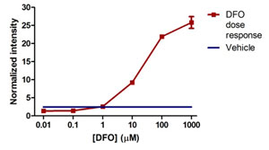
Sample experiment using ab125299 on HeLa cells treated with a titration of DFO. HeLa cells were seeded to an amine coated 96-well microplate and the following day treated with a titration of DFO. After 24h of DFO exposure, the cells were fixed and stained as described in the protocol and the normalized data is presented here +/- SD (as described in the protocol and data analysis sections). HIF1 alpha results show DFO concentrations >10µM induce HIF1A protein levels in a dose dependent fashion.
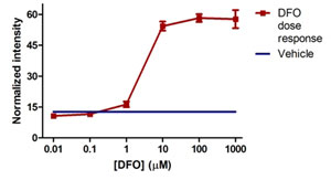
Sample experiment using ab125299 on HeLa cells treated with a titration of DFO. HeLa cells were seeded to an amine coated 96-well microplate and the following day treated with a titration of DFO. After 24h of DFO exposure, the cells were fixed and stained as described in the protocol and the normalized data is presented here +/- SD (as described in the protocol and data analysis sections). PDK1 levels are increased with >10µM DFO.
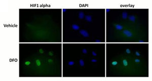
Antibody specificity demonstrated by immunocytochemistry. Primary antibodies used in this assay kit were validated by staining HeLa cells +/- treatment with 1mM DFO (24h) and imaged by fluorescent microscopy. HIF1 alpha (ab51608, diluted 1:2000) staining is absent in untreated cells and induced by DFO treatment. HIF1 alpha localizes to the nucleus (as seen by co-localization with the DNA stain DAPI) as expected.
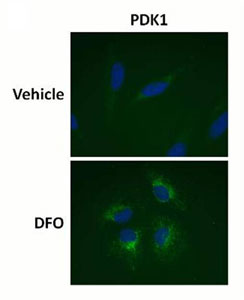
Antibody specificity demonstrated by immunocytochemistry. Primary antibodies used in this assay kit were validated by staining HeLa cells +/- treatment with 1mM DFO (24h) and imaged by fluorescent microscopy. PDK1 staining (ab110335, diluted 2µg/mL) labels cellular mitochondria and the fluorescent intensity increases with DFO treatment.
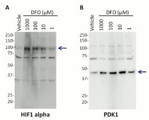
Antibody specificity demonstrated by western blot. Primary antibodies used in this assay kit were validated by western blot using HeLa cell lysates that had been treated with a dose titration of DFO as indicated. (A) The HIF1 alpha (ab51608) band (indicated by arrow) is absent in untreated cells and induced in a dose-dependent manner by DFO. (B) Similarly, PDK1 levels are increased by DFO treatment in a dose-dependent manner.
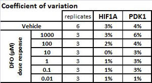
Coefficient of variation for the experiment described in Figure 1.