![All lanes : Anti-Cytochrome C antibody [EPR1327] (HRP) (ab193239) at 1/5000 dilutionLane 1 : SHSY-5Y (Human neuroblastoma cell line) Whole Cell LysateLane 2 : Heart (Human) Tissue Lysate - adult normal tissueLane 3 : Kidney (Human) Tissue Lysate - adult normal tissueLysates/proteins at 10 µg per lane.developed using the ECL techniquePerformed under reducing conditions.](http://www.bioprodhub.com/system/product_images/ab_products/2/sub_2/4727_ab193239-234243-WBab1932391.jpg)
All lanes : Anti-Cytochrome C antibody [EPR1327] (HRP) (ab193239) at 1/5000 dilutionLane 1 : SHSY-5Y (Human neuroblastoma cell line) Whole Cell LysateLane 2 : Heart (Human) Tissue Lysate - adult normal tissueLane 3 : Kidney (Human) Tissue Lysate - adult normal tissueLysates/proteins at 10 µg per lane.developed using the ECL techniquePerformed under reducing conditions.
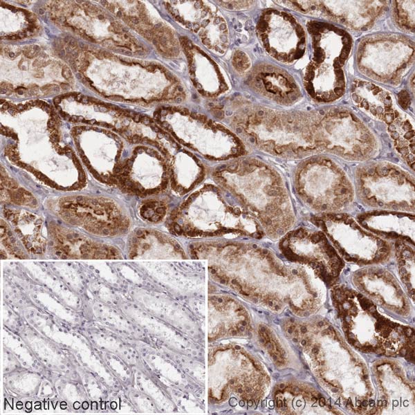
IHC image of Cytochrome C staining in a section of formalin-fixed paraffin-embedded normal Human kidney tissue. The section was pre-treated using pressure cooker heat mediated antigen retrieval with sodium citrate buffer (pH6) for 30 mins. The section was incubated with ab193239 at a working dilution of 1 in 10000 overnight at +4°C. The section was counterstained with haematoxylin and mounted with DPX.The inset negative control image is taken from an identical assay without primary antibody.For other IHC staining systems (automated and non-automated) customers should optimize variable parameters such as antigen retrieval conditions, primary antibody concentration and antibody incubation times.*Tissue obtained from the Human Research Tissue Bank, supported by the NIHR Cambridge Biomedical Research Centre.