Anti-Cytochrome C antibody [EPR1327]
| Name | Anti-Cytochrome C antibody [EPR1327] |
|---|---|
| Supplier | Abcam |
| Catalog | ab133504 |
| Prices | $387.00 |
| Sizes | 100 µl |
| Host | Rabbit |
| Clonality | Monoclonal |
| Isotype | IgG |
| Clone | EPR1327 |
| Applications | WB IHC-P ICC/IF ICC/IF IP |
| Species Reactivities | Mouse, Rat, Human |
| Antigen | Synthetic peptide corresponding to residues in Human Cytochrome C |
| Description | Rabbit Monoclonal |
| Gene | CYCS |
| Conjugate | Unconjugated |
| Supplier Page | Shop |
Product images
![Anti-Cytochrome C antibody [EPR1327] (ab133504) at 1/5000 dilution (purified) + Human fetal kidney tissue lysate at 10 µgSecondaryHRP goat anti-rabbit (H+L) at 1/1000 dilution](http://www.bioprodhub.com/system/product_images/ab_products/2/sub_2/4711_ab133504-241442-133504-WB-3.jpg) Anti-Cytochrome C antibody [EPR1327] (ab133504) at 1/5000 dilution (purified) + Human fetal kidney tissue lysate at 10 µgSecondaryHRP goat anti-rabbit (H+L) at 1/1000 dilution
Anti-Cytochrome C antibody [EPR1327] (ab133504) at 1/5000 dilution (purified) + Human fetal kidney tissue lysate at 10 µgSecondaryHRP goat anti-rabbit (H+L) at 1/1000 dilution
![Anti-Cytochrome C antibody [EPR1327] (ab133504) at 1/5000 dilution (purified) + Human fetal heart tissue lysate at 10 µgSecondaryHRP goat anti-rabbit (H+L) at 1/1000 dilution](http://www.bioprodhub.com/system/product_images/ab_products/2/sub_2/4712_ab133504-241441-133504-WB-4.jpg) Anti-Cytochrome C antibody [EPR1327] (ab133504) at 1/5000 dilution (purified) + Human fetal heart tissue lysate at 10 µgSecondaryHRP goat anti-rabbit (H+L) at 1/1000 dilution
Anti-Cytochrome C antibody [EPR1327] (ab133504) at 1/5000 dilution (purified) + Human fetal heart tissue lysate at 10 µgSecondaryHRP goat anti-rabbit (H+L) at 1/1000 dilution
![Anti-Cytochrome C antibody [EPR1327] (ab133504) at 1/5000 dilution (purified) + Rat brain tissue lysate at 1/1000 dilutionSecondaryHRP goat anti-rabbit (H+L) at 1/1000 dilution](http://www.bioprodhub.com/system/product_images/ab_products/2/sub_2/4713_ab133504-241440-133504-WB-1.jpg) Anti-Cytochrome C antibody [EPR1327] (ab133504) at 1/5000 dilution (purified) + Rat brain tissue lysate at 1/1000 dilutionSecondaryHRP goat anti-rabbit (H+L) at 1/1000 dilution
Anti-Cytochrome C antibody [EPR1327] (ab133504) at 1/5000 dilution (purified) + Rat brain tissue lysate at 1/1000 dilutionSecondaryHRP goat anti-rabbit (H+L) at 1/1000 dilution
![Anti-Cytochrome C antibody [EPR1327] (ab133504) at 1/50000 dilution (purified) + Mouse brain tissue lysate at 10 µgSecondaryHRP goat anti-rabbit (H+L) at 1/1000 dilution](http://www.bioprodhub.com/system/product_images/ab_products/2/sub_2/4714_ab133504-241439-133504-WB-2.jpg) Anti-Cytochrome C antibody [EPR1327] (ab133504) at 1/50000 dilution (purified) + Mouse brain tissue lysate at 10 µgSecondaryHRP goat anti-rabbit (H+L) at 1/1000 dilution
Anti-Cytochrome C antibody [EPR1327] (ab133504) at 1/50000 dilution (purified) + Mouse brain tissue lysate at 10 µgSecondaryHRP goat anti-rabbit (H+L) at 1/1000 dilution
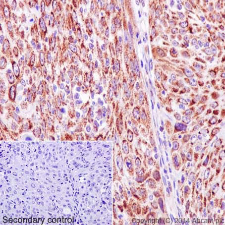 Immunohistochemical staining of paraffin embedded human cervical carcinoma with purified ab133504 at a working dilution of 1 in 500. The secondary antibody used is a HRP goat anti-rabbit H+L (ab97051). The sample is counter-stained with hematoxylin. Antigen retrieval was perfomed using Tris-EDTA buffer, pH 9.0. PBS was used instead of the primary antibody as the negative control, and is shown in the inset.
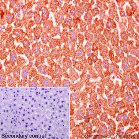 Immunohistochemical staining of paraffin embedded mouse liver with purified ab133504 at a working dilution of 1 in 500. The secondary antibody used is a HRP goat anti-rabbit H+L (ab97051). The sample is counter-stained with hematoxylin. Antigen retrieval was perfomed using Tris-EDTA buffer, pH 9.0. PBS was used instead of the primary antibody as the negative control, and is shown in the inset.
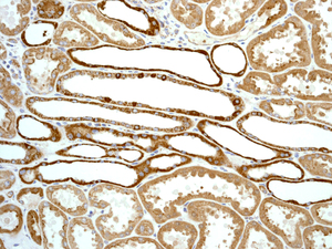
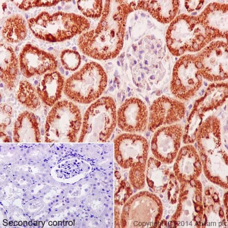 Immunohistochemical staining of paraffin embedded rat kidney with purified ab133504 at a working dilution of 1 in 500. The secondary antibody used is a HRP goat anti-rabbit H+L (ab97051). The sample is counter-stained with hematoxylin. Antigen retrieval was perfomed using Tris-EDTA buffer, pH 9.0. PBS was used instead of the primary antibody as the negative control, and is shown in the inset.
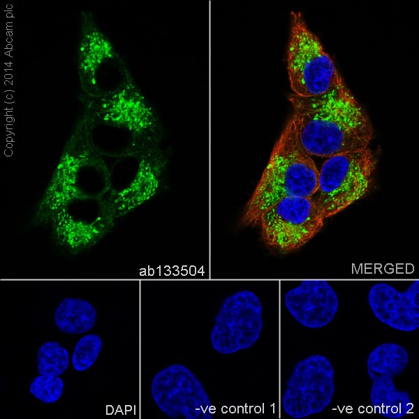 Immunofluorescence staining of SH-SY5Y cells with purified ab133504 at a working dilution of 1 in 100, counter-stained with DAPI. The secondary antibody was Alexa Fluor® 488 goat anti rabbit (ab150077), used at a dilution of 1 in 500. ab7291 was used to stain tubulin, and this is shown in the top right hand panel. The cells were fixed in 4% PFA and permeabilized using 0.1% Triton X 100. The negative control is shown in bottom middle and right hand panels - for the negative controls, purified ab133504 was used at a dilution of 1/200 followed by an Alexa Fluor® 594 goat anti-mouse antibody at a dilution of 1/500.
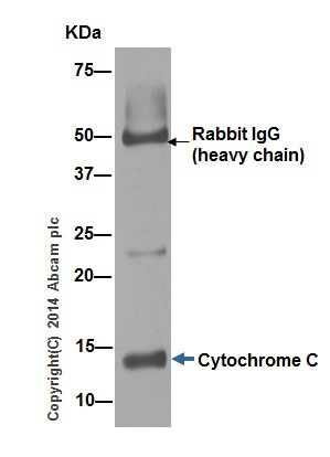 ab133504 (purified) at 1/30 immunoprecipitating Cytochrome C in Molt-4 cells (Lane 1). For western blotting, a HRP-conjugated goat anti-rabbit (H+L), was used as the secondary antibody (1/1000).Blocking buffer and concentration: 5% NFDM/TBST.Diluting buffer and concentration: 5% NFDM /TBST.
![All lanes : Anti-Cytochrome C antibody [EPR1327] (ab133504) at 1/10000 dilution (unpurified)Lane 1 : Molt4 lysateLane 2 : SH-SY5Y lysateLane 3 : Human heart lysateLane 4 : Human kidney lysateLane 5 : Human spleen lysate Lysates/proteins at 10 µg per lane.SecondaryGoat-anti-rabbit HRP at 1/2000 dilution](http://www.bioprodhub.com/system/product_images/ab_products/2/sub_2/4720_Cytochrome-C-Primary-antibodies-ab133504-3.jpg) All lanes : Anti-Cytochrome C antibody [EPR1327] (ab133504) at 1/10000 dilution (unpurified)Lane 1 : Molt4 lysateLane 2 : SH-SY5Y lysateLane 3 : Human heart lysateLane 4 : Human kidney lysateLane 5 : Human spleen lysate Lysates/proteins at 10 µg per lane.SecondaryGoat-anti-rabbit HRP at 1/2000 dilution
All lanes : Anti-Cytochrome C antibody [EPR1327] (ab133504) at 1/10000 dilution (unpurified)Lane 1 : Molt4 lysateLane 2 : SH-SY5Y lysateLane 3 : Human heart lysateLane 4 : Human kidney lysateLane 5 : Human spleen lysate Lysates/proteins at 10 µg per lane.SecondaryGoat-anti-rabbit HRP at 1/2000 dilution
 Equilibrium disassociation constant (KD)Learn more about KD Click here to learn more about KD
 Immunohistochemical analysis of paraffin-embedded Human kidney tissue labelling Cytochrome C with unpurified ab133504 at 1/250 dilution.
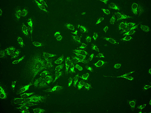 Immunofluorescent analysis of HeLa cells labelling Cytochrome C with unpurified ab133504 at 1/100 dilution.
Product References
The protease Omi regulates mitochondrial biogenesis through the - The protease Omi regulates mitochondrial biogenesis through the
Xu R, Hu Q, Ma Q, Liu C, Wang G. Cell Death Dis. 2014 Aug 14;5:e1373.
Enhancer of zeste homolog 2 is a negative regulator of mitochondria-mediated - Enhancer of zeste homolog 2 is a negative regulator of mitochondria-mediated
Chen S, Sheng C, Liu D, Yao C, Gao S, Song L, Jiang W, Li J, Huang W. J Immunol. 2013 Sep 1;191(5):2614-23.