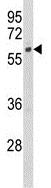
Anti-CYP7B1 antibody (ab175889) at 1/1000 dilution + HeLa cell lysate at 35 µg
Immunohistochemical analysis of Formalin-fixed, Paraffin-embedded Human brain tissue labeling CYP7B1 with ab175889 at 1/50 dilution followed by peroxidase-conjugated secondary antibody and DAB staining.
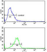
Flow Cytometric analysis of HeLa cells labeling CYP7B1 with ab175889 at 1/10 dilution (bottom histogram) compared to a negative control cell (top histogram). FITC-conjugated goat-anti-rabbit secondary antibodies were used for the analysis.
Immunofluorescent analysis of HeLa cells labeling CYP7B1 with ab175889 at 1/10 dilution followed by Alexa Fluor® 488-conjugated goat anti-rabbit lgG (green). DAPI was used to stain the cell nuclear (blue).