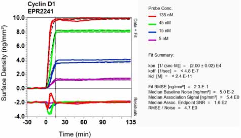
Anti-Cyclin D1 antibody [EPR2241]
| Name | Anti-Cyclin D1 antibody [EPR2241] |
|---|---|
| Supplier | Abcam |
| Catalog | ab134175 |
| Prices | $400.00 |
| Sizes | 100 µl |
| Host | Rabbit |
| Clonality | Monoclonal |
| Isotype | IgG |
| Clone | EPR2241 |
| Applications | WB IP ICC/IF ICC/IF IHC-P |
| Species Reactivities | Mouse, Rat, Human |
| Antigen | Synthetic peptide (the amino acid sequence is considered to be commercially sensitive) corresponding to Human Cyclin D1 (C terminal) |
| Blocking Peptide | Cyclin D1 peptide |
| Description | Rabbit Monoclonal |
| Gene | CCND1 |
| Conjugate | Unconjugated |
| Supplier Page | Shop |
Product images
Product References
Elevated expression of TANK-binding kinase 1 enhances tamoxifen resistance in - Elevated expression of TANK-binding kinase 1 enhances tamoxifen resistance in
Wei C, Cao Y, Yang X, Zheng Z, Guan K, Wang Q, Tai Y, Zhang Y, Ma S, Cao Y, Ge X, Xu C, Li J, Yan H, Ling Y, Song T, Zhu L, Zhang B, Xu Q, Hu C, Bian XW, He X, Zhong H. Proc Natl Acad Sci U S A. 2014 Feb 4;111(5):E601-10. doi:
Overexpression of Cdk6 and Ccnd1 in chondrocytes inhibited chondrocyte maturation - Overexpression of Cdk6 and Ccnd1 in chondrocytes inhibited chondrocyte maturation
Ito K, Maruyama Z, Sakai A, Izumi S, Moriishi T, Yoshida CA, Miyazaki T, Komori H, Takada K, Kawaguchi H, Komori T. Oncogene. 2014 Apr 3;33(14):1862-71.
Elevated cyclin D1 expression is predictive for a benefit from TPF induction - Elevated cyclin D1 expression is predictive for a benefit from TPF induction
Zhong LP, Zhu DW, William WN Jr, Liu Y, Ma J, Yang CZ, Yang X, Wang LZ, Li J, Myers JN, Lee JJ, Zhang CP, Zhang ZY. Mol Cancer Ther. 2013 Jun;12(6):1112-21.
Akt-p53-miR-365-cyclin D1/cdc25A axis contributes to gastric tumorigenesis - Akt-p53-miR-365-cyclin D1/cdc25A axis contributes to gastric tumorigenesis
Guo SL, Ye H, Teng Y, Wang YL, Yang G, Li XB, Zhang C, Yang X, Yang ZZ, Yang X. Nat Commun. 2013;4:2544.
Molecular chaperone gp96 is a novel therapeutic target of multiple myeloma. - Molecular chaperone gp96 is a novel therapeutic target of multiple myeloma.
Hua Y, White-Gilbertson S, Kellner J, Rachidi S, Usmani SZ, Chiosis G, Depinho R, Li Z, Liu B. Clin Cancer Res. 2013 Nov 15;19(22):6242-51.
The cannabinoid WIN 55,212-2 decreases specificity protein transcription factors - The cannabinoid WIN 55,212-2 decreases specificity protein transcription factors
Sreevalsan S, Safe S. Mol Cancer Ther. 2013 Nov;12(11):2483-93.
CCND1 as a predictive biomarker of neoadjuvant chemotherapy in patients with - CCND1 as a predictive biomarker of neoadjuvant chemotherapy in patients with
Feng Z, Guo W, Zhang C, Xu Q, Zhang P, Sun J, Zhu H, Wang Z, Li J, Wang L, Wang B, Ren G, Ji T, Tu W, Yang X, Qiu W, Mao L, Zhang Z, Chen W. PLoS One. 2011;6(10):e26399.
![All lanes : Anti-Cyclin D1 antibody [EPR2241] (ab134175) at 1/10000 dilution (purified)Lane 1 : Mouse kidneyLane 2 : Mouse spleenLane 3 : Rat heartLysates/proteins at 20 µg per lane.SecondaryHRP goat anti-rabbit (H+L) at 1/1000 dilution](http://www.bioprodhub.com/system/product_images/ab_products/2/sub_2/3949_ab134175-237377-134175-WB-3.jpg)
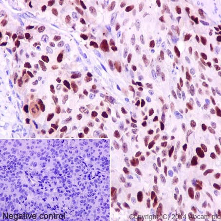
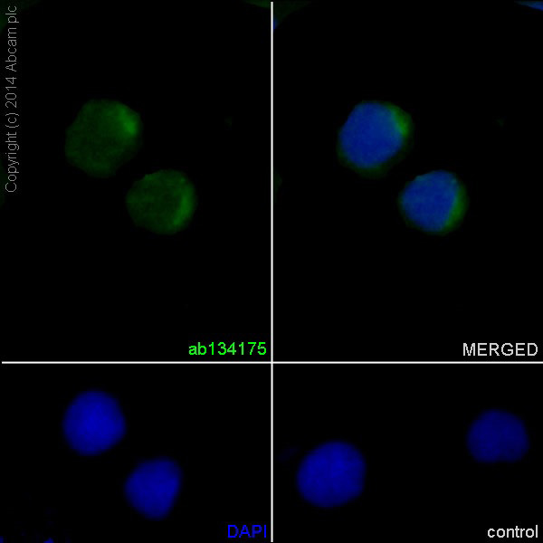
![All lanes : Anti-Cyclin D1 antibody [EPR2241] (ab134175) at 1/100000 dilution (purified)Lane 1 : MCF-7 cell lysateLane 2 : LnCap cell lysateLysates/proteins at 20 µg per lane.SecondaryHRP goat anti-rabbit (H+L) at 1/1000 dilution](http://www.bioprodhub.com/system/product_images/ab_products/2/sub_2/3952_ab134175-237376-134175-WB-2.jpg)
![All lanes : Anti-Cyclin D1 antibody [EPR2241] (ab134175) at 1/20000 dilution (purified)Lane 1 : HeLa cell lysateLane 2 : A431 cell lysateLysates/proteins at 20 µg per lane.SecondaryHRP goat anti-rabbit (H+L) at 1/1000 dilution](http://www.bioprodhub.com/system/product_images/ab_products/2/sub_2/3953_ab134175-237375-134175-WB-1.jpg)
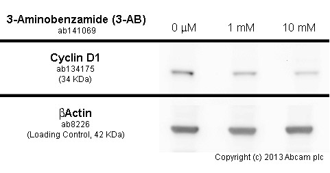
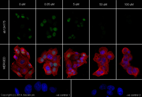
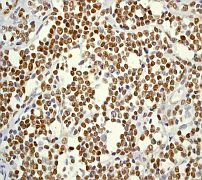
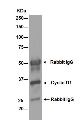
![Anti-Cyclin D1 antibody [EPR2241] (ab134175) at 1/10000 dilution (unpurified) + MCF7 cell lysate at 10 µgSecondaryHRP labelled goat anti-rabbit at 1/2000 dilution](http://www.bioprodhub.com/system/product_images/ab_products/2/sub_2/3958_Cyclin-D1-Primary-antibodies-ab134175-3.jpg)
