![Anti-CTPS2 antibody [EPR16735] (ab196016) at 1/20000 dilution + HeLa (Human epithelial cells from cervix adenocarcinoma) cell lysate at 20 µgSecondaryGoat Anti-Rabbit IgG, (H+L), Peroxidase conjugated at 1/1000 dilution](http://www.bioprodhub.com/system/product_images/ab_products/2/sub_2/3220_ab196016-241801-ab196016-wb-1.jpg)
Anti-CTPS2 antibody [EPR16735] (ab196016) at 1/20000 dilution + HeLa (Human epithelial cells from cervix adenocarcinoma) cell lysate at 20 µgSecondaryGoat Anti-Rabbit IgG, (H+L), Peroxidase conjugated at 1/1000 dilution
![All lanes : Anti-CTPS2 antibody [EPR16735] (ab196016) at 1/2000 dilutionLane 1 : HepG2 (Human liver hepatocellular carcinoma) cell lysateLane 2 : 293 (Human epithelial cells from embryonic kidney) cell lysateLysates/proteins at 20 µg per lane.SecondaryGoat Anti-Rabbit IgG, (H+L), Peroxidase conjugated at 1/1000 dilution](http://www.bioprodhub.com/system/product_images/ab_products/2/sub_2/3221_ab196016-241800-ab196016-wb-2.jpg)
All lanes : Anti-CTPS2 antibody [EPR16735] (ab196016) at 1/2000 dilutionLane 1 : HepG2 (Human liver hepatocellular carcinoma) cell lysateLane 2 : 293 (Human epithelial cells from embryonic kidney) cell lysateLysates/proteins at 20 µg per lane.SecondaryGoat Anti-Rabbit IgG, (H+L), Peroxidase conjugated at 1/1000 dilution
![All lanes : Anti-CTPS2 antibody [EPR16735] (ab196016) at 1/2000 dilutionLane 1 : Raw264.7 (Mouse macrophage cells transformed with Abelson murine leukemia virus) cell lysateLane 2 : PC12 (Rat adrenal gland pheochromocytoma) cell lysateLysates/proteins at 10 µg per lane.SecondaryGoat Anti-Rabbit IgG, (H+L), Peroxidase conjugated at 1/1000 dilution](http://www.bioprodhub.com/system/product_images/ab_products/2/sub_2/3222_ab196016-241799-ab196016-wb-3.jpg)
All lanes : Anti-CTPS2 antibody [EPR16735] (ab196016) at 1/2000 dilutionLane 1 : Raw264.7 (Mouse macrophage cells transformed with Abelson murine leukemia virus) cell lysateLane 2 : PC12 (Rat adrenal gland pheochromocytoma) cell lysateLysates/proteins at 10 µg per lane.SecondaryGoat Anti-Rabbit IgG, (H+L), Peroxidase conjugated at 1/1000 dilution
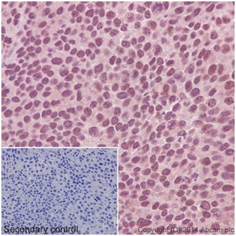
Immunohistochemical analysis of paraffin-embedded Human transitional cell carcinoma of bladder tissue labeling CTPS2 using ab196016 at 1/1500 dilution, followed by Goat Anti-Rabbit IgG H&L (HRP) (ab97051) at 1/500 dilution. Cytoplasmic and Nuclear staining is observed. Counter stained with Hematoxylin. Negative control: Used PBS instead of primary antibody, secondary antibody is Goat Anti-Rabbit IgG H&L (HRP) (ab97051) at 1/500 dilution.The staining pattern observed is consistent with what has been described in the literature PMID: 24870241 and PMID: 16179339.
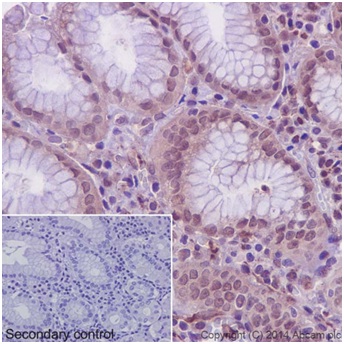
Immunohistochemical analysis of paraffin-embedded Human stomach tissue labeling CTPS2 using ab196016 at 1/1500 dilution, followed by Goat Anti-Rabbit IgG H&L (HRP) (ab97051) at 1/500 dilution. Cytoplasmic and Nuclear staining is observed. Counter stained with Hematoxylin.Negative control: Used PBS instead of primary antibody, secondary antibody is Goat Anti-Rabbit IgG H&L (HRP) (ab97051) at 1/500 dilution.The staining pattern observed is consistent with what has been described in the literature PMID: 24870241 and PMID: 16179339.
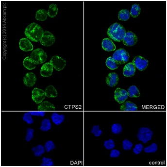
Immunofluorescent analysis of 4% paraformaldehyde-fixed, 0.1% Triton X-100 permeabilized K562 (Human chronic myelogenous leukemia cells from bone marrow) cells labeling CTPS2 with ab196016 at 1/600 dilution, followed by Goat anti-rabbit IgG (Alexa Fluor® 488) (ab150077) secondary antibody at 1/500 dilution (green). Cytoplasmic and Nuclear staining is observed. The nuclear counter stain is DAPI (blue).Negative control obtained using PBS instead of ab196016 followed by Goat anti-rabbit IgG (Alexa Fluor® 488) (ab150077) secondary antibody at 1/500 dilution.The staining pattern observed is consistent with what has been described in the literature PMID: 16179339 and 24870241.
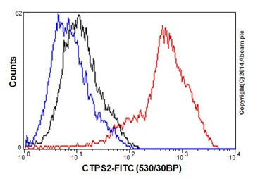
Flow cytometry analysis of HeLa (Human epithelial cells from cervix adenocarcinoma) cells labeling CTPS2 using ab196016 at 1/540 dilution (Red). A Goat anti rabbit IgG (FITC) at 1/150 dilution was used as secondary antibody. Cells were fixed with 2% paraformaldehyde. Cells without incubation with primary antibody and secondary antibody (Blue). Rabbit monoclonal IgG was used as isotype control (Black).