![All lanes : Anti-Ctip1 [EPR14943-2] antibody (ab191402) at 1/20000 dilutionLane 1 : Raji cell lysateLane 2 : Jurkat cell lysateLane 3 : 293 cell lysateLysates/proteins at 20 µg per lane.Secondarygoat anti-rabbit IgG, (H+L), peroxidase conjugated at 1/1000 dilutiondeveloped using the ECL technique](http://www.bioprodhub.com/system/product_images/ab_products/2/sub_2/3077_ab191402-228678-ab191402WB.jpg)
All lanes : Anti-Ctip1 [EPR14943-2] antibody (ab191402) at 1/20000 dilutionLane 1 : Raji cell lysateLane 2 : Jurkat cell lysateLane 3 : 293 cell lysateLysates/proteins at 20 µg per lane.Secondarygoat anti-rabbit IgG, (H+L), peroxidase conjugated at 1/1000 dilutiondeveloped using the ECL technique
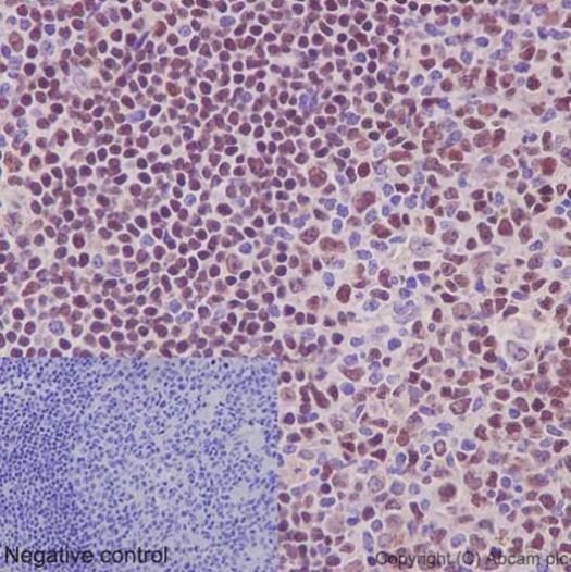
Immunohistochemical analysis of paraffin-embedded, Human tonsil tissue labeling Ctip1 with ab191402 at a 1/100 dilution (23.5 μg/ml). Counter stained with hematoxylin. In the negative control PBS was used instead of primary antibody.
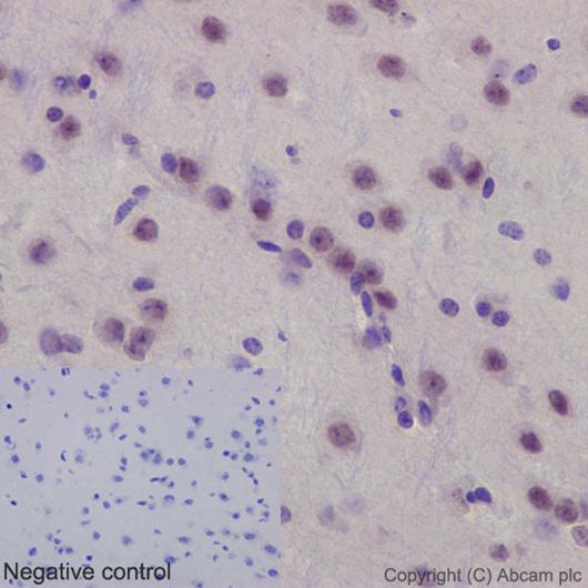
Immunohistochemical analysis of paraffin-embedded, mouse brain tissue labeling Ctip1 with ab191402 at a 1/100 dilution (23.5 μg/ml). Counter stained with hematoxylin. In the negative control PBS was used instead of primary antibody.
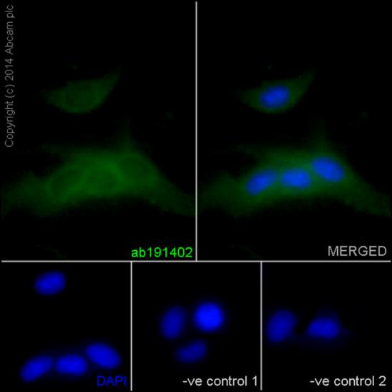
Immunofluorescence analysis of, paraformaldehyde-fixed, rat C6 cells labeling Ctip1 with ab191402 at a 1/600 dilution (4 ug/ml). As secondary antibody goat anti-rabbit IgG (Alexa Fluor®488) ab150077 was used at a 1/200 dilution. In blue DAPI staining. In the negative controls cells were treated with anti-Ctip1 at a 1/600 dilution as primary antibody and goat anti-mouse IgG (Alexa Fluor®594) at a 1/400 dilution as secondary antibody.
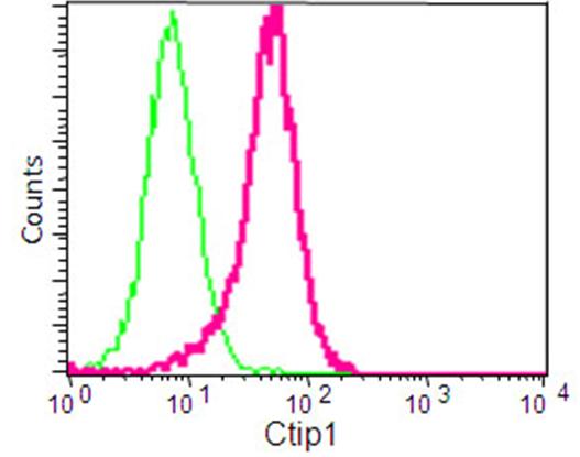
Flow cytometry analysis of paraformaldehyde-fixed 293 cells labeling Ctip1 with ab191402 at a 1/250 dilution and secondary antibody goat anti-rabbit IgG (FITC, red) at a 1/150 dilution, or negative control rabbit IgG (green).