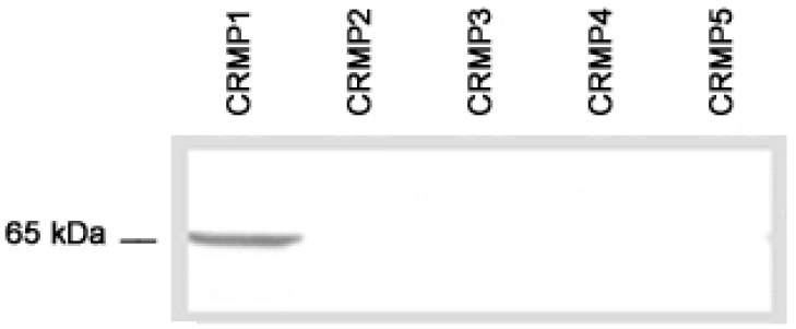
All lanes : Anti-CRMP1 antibody (ab36199)Lane 1 : E.coli lysate transformed with cDNA of human CRMP1.Lane 2 : E.coli lysate transformed with cDNA of human CRMP2.Lane 3 : E.coli lysate transformed with cDNA of human CRMP3.Lane 4 : E.coli lysate transformed with cDNA of human CRMP4.Lane 5 : E.coli lysate transformed with cDNA of human CRMP5.

Ab36199 staining human CRMP1 in olfactory receptor neurons within the olfactory mucosa by immunohistochemistry.
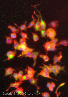
ICC/IF image of ab36199 stained PC12 cells. The cells were 4% formaldehyde fixed (10 min) and then incubated in 1%BSA / 10% normal goat serum / 0.3M glycine in 0.1% PBS-Tween for 1h to permeabilise the cells and block non-specific protein-protein interactions. The cells were then incubated with the antibody (ab36199, 1/1000 dilution) overnight at +4°C. The secondary antibody (green) was Alexa Fluor® 488 goat anti-rabbit IgG (H+L) used at a 1/1000 dilution for 1h. Alexa Fluor® 594 WGA was used to label plasma membranes (red) at a 1/200 dilution for 1h. DAPI was used to stain the cell nuclei (blue) at a concentration of 1.43µM.