Anti-CRIP1 antibody - C-terminal
| Name | Anti-CRIP1 antibody - C-terminal |
|---|---|
| Supplier | Abcam |
| Catalog | ab174325 |
| Prices | $370.00 |
| Sizes | 400 µl |
| Host | Rabbit |
| Clonality | Polyclonal |
| Isotype | IgG |
| Applications | WB FC IHC-P |
| Species Reactivities | Mouse, Human, Rat, Bovine |
| Antigen | Synthetic peptide within Human CRIP1 aa 48-77 (C terminal) conjugated to Keyhole Limpet Haemocyanin (KLH) |
| Description | Rabbit Polyclonal |
| Gene | CRIP1 |
| Conjugate | Unconjugated |
| Supplier Page | Shop |
Product images
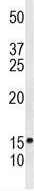 Anti-CRIP1 antibody - C-terminal (ab174325) at 1/100 dilution + HL60 cell line lysate at 35 µg
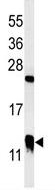 Anti-CRIP1 antibody - C-terminal (ab174325) at 1/100 dilution + Mouse bladder tissue lysate at 35 µg
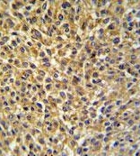 Immunohistochemical analysis of formalin fixed and paraffin embedded bladder carcinoma tissue labeling CRIP1 with ab174325 at 1/50 followed by peroxidase conjugation of the secondary antibody and DAB staining.
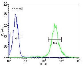 Flow Cytometrical analysis of HL60 cells with ab174325 antibody at a dilution of 1/10 (right histogram) compared to a negative control cell (left histogram). FITC-conjugated goat-anti-rabbit secondary antibodies were used for the analysis.