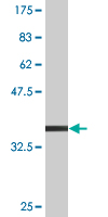
Anti-Creatine kinase MT antibody (ab76506) at 1 µg/ml + Recombinant tagged human Creatine kinase MT fragment at 0.1 µgSecondaryGoat Anti-Mouse IgG (H&L)-HRP Conjugate at 1/5000 dilution
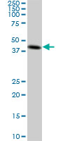
Anti-Creatine kinase MT antibody (ab76506) at 1 µg/ml + A-431 cell lysate at 25 µgSecondaryGoat Anti-Mouse IgG (H&L)-HRP Conjugate at 1/2500 dilution
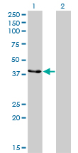
All lanes : Anti-Creatine kinase MT antibody (ab76506) at 1 µg/mlLane 1 : Creatine kinase MT transfected 293T cell lysateLane 2 : Non-transfected 293T cell lysateLysates/proteins at 25 µg per lane.SecondaryGoat Anti-Mouse IgG (H&L)-HRP Conjugate at 1/2500 dilution
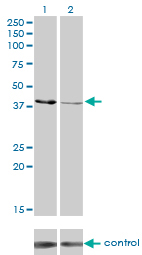
All lanes : Anti-Creatine kinase MT antibody (ab76506) at 1 µg/mlLane 1 : Creatine kinase MT over-expressed 293 cell line, non-transfected control Lane 2 : Creatine kinase MT over-expressed 293 cell line, cotransfected with Creatine kinase MT Validated Chimera RNAi Lysates/proteins at 25 µg per lane.SecondaryGoat Anti-Mouse IgG (H&L)-HRP Conjugate at 1/2500 dilution
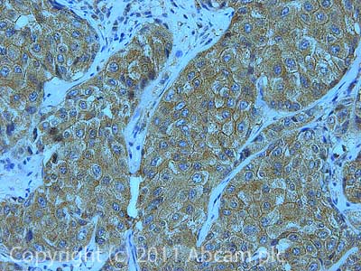
IHC image of ab76506 staining in Human Breast cancer formalin fixed paraffin embedded tissue section, performed on a Leica BondTM system using the standard protocol F. The section was pre-treated using heat mediated antigen retrieval with sodium citrate buffer (pH6, epitope retrieval solution 1) for 20 mins. The section was then incubated with ab76506, 1µg/ml, for 15 mins at room temperature and detected using an HRP conjugated compact polymer system. DAB was used as the chromogen. The section was then counterstained with haematoxylin and mounted with DPX.For other IHC staining systems (automated and non-automated) customers should optimize variable parameters such as antigen retrieval conditions, primary antibody concentration and antibody incubation times.
![Overlay histogram showing HeLa cells stained with ab76506 (red line). The cells were fixed with 80% methanol (5 min) and then permeabilized with 0.1% PBS-Tween for 20 min. The cells were then incubated in 1x PBS / 10% normal goat serum / 0.3M glycine to block non-specific protein-protein interactions followed by the antibody (ab76506, 0.5µg/1x106 cells) for 30 min at 22ºC. The secondary antibody used was DyLight® 488 goat anti-mouse IgG (H+L) (ab96879) at 1/500 dilution for 30 min at 22ºC. Isotype control antibody (black line) was mouse IgG2a [ICIGG2A] (ab91361, 1µg/1x106 cells) used under the same conditions. Acquisition of >5,000 events was performed. This antibody gave a positive signal in HeLa cells fixed with 4% paraformaldehyde (10 min)/permeabilized with 0.1% PBS-Tween for 20 min used under the same conditions.](http://www.bioprodhub.com/system/product_images/ab_products/2/sub_2/2065_Creatine-kinase-MT-Primary-antibodies-ab76506-12.jpg)
Overlay histogram showing HeLa cells stained with ab76506 (red line). The cells were fixed with 80% methanol (5 min) and then permeabilized with 0.1% PBS-Tween for 20 min. The cells were then incubated in 1x PBS / 10% normal goat serum / 0.3M glycine to block non-specific protein-protein interactions followed by the antibody (ab76506, 0.5µg/1x106 cells) for 30 min at 22ºC. The secondary antibody used was DyLight® 488 goat anti-mouse IgG (H+L) (ab96879) at 1/500 dilution for 30 min at 22ºC. Isotype control antibody (black line) was mouse IgG2a [ICIGG2A] (ab91361, 1µg/1x106 cells) used under the same conditions. Acquisition of >5,000 events was performed. This antibody gave a positive signal in HeLa cells fixed with 4% paraformaldehyde (10 min)/permeabilized with 0.1% PBS-Tween for 20 min used under the same conditions.