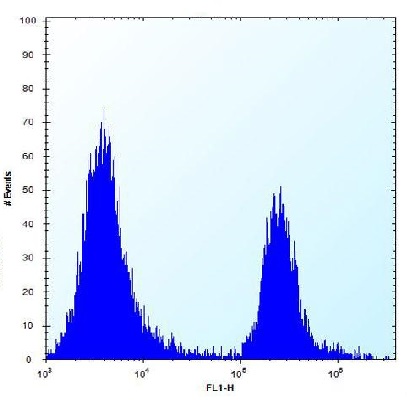
Flow cytometric analysis of 293 cells (right histogram) compared to a negative control cell (left histogram). An FITC-conjugated goat-anti-rabbit secondary antibody was used for the analysis.
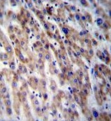
Immunohistochemical analysis of formalin fixed and paraffin embedded Human liver tissue labeling CPN1, using ab171323 at a 1/10 dilution, followed by peroxidase conjugation of the secondary antibody and DAB staining.
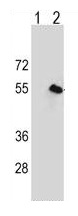
All lanes : Anti-CPN1 antibody (ab171323) at 1/50 dilutionLane 1 : 293 cell lysateLane 2 : 293 cell lysate transiently transfected with the CPN1 geneLysates/proteins at 2 µg per lane.