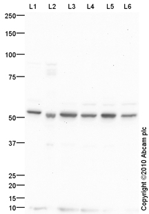
All lanes : Anti-Choline kinase alpha antibody (ab88053) at 1 µg/mlLane 1 : MCF7 (Human breast adenocarcinoma cell line) Whole Cell LysateLane 2 : Breast (Human) Tissue Lysate - normal tissue (ab30090)Lane 3 : HepG2 (Human hepatocellular liver carcinoma cell line) Whole Cell LysateLane 4 : HeLa (Human epithelial carcinoma cell line) Whole Cell LysateLane 5 : Jurkat (Human T cell lymphoblast-like cell line) Whole Cell LysateLane 6 : A549 (Human lung adenocarcinoma epithelial cell line) Whole Cell Lysate Lysates/proteins at 10 µg per lane.SecondaryGoat polyclonal to Rabbit IgG - H&L - Pre-Adsorbed (HRP) at 1/3000 dilutiondeveloped using the ECL techniquePerformed under reducing conditions.
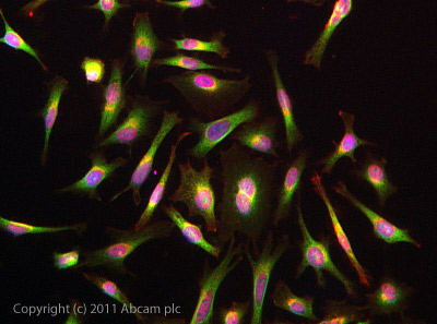
ICC/IF image of ab88053 stained HeLa cells. The cells were 4% PFA fixed (10 min) and then incubated in 1%BSA / 10% normal goat serum / 0.3M glycine in 0.1% PBS-Tween for 1h to permeabilise the cells and block non-specific protein-protein interactions. The cells were then incubated with the antibody (ab88053, 5µg/ml) overnight at +4°C. The secondary antibody (green) was ab96899 Dylight 488 goat anti-rabbit IgG (H+L) used at a 1/250 dilution for 1h. Alexa Fluor® 594 WGA was used to label plasma membranes (red) at a 1/200 dilution for 1h. DAPI was used to stain the cell nuclei (blue) at a concentration of 1.43µM.
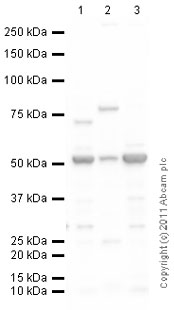
All lanes : Anti-Choline kinase alpha antibody (ab88053) at 1 µg/mlLane 1 : Brain (Mouse) Tissue LysateLane 2 : Lung (Mouse) Whole Cell Lysate - normal tissue (ab29297)Lane 3 : Brain (Rat) Tissue LysateLysates/proteins at 10 µg per lane.SecondaryGoat Anti-Rabbit IgG H&L (HRP) preadsorbed (ab97080) at 1/5000 dilutiondeveloped using the ECL techniquePerformed under reducing conditions.