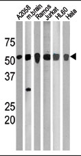
All lanes : Anti-Chk1 (phospho S280) antibody (ab59988) at 1/100 dilutionLane 1 : A2058 cell lysateLane 2 : mouse brain lysateLane 3 : Ramos cell lysateLane 4 : Jurkat cell lysateLane 5 : HL60 cell lysate Lane 6 : Hela cell lysate
ab59988 at 1/50-1/100 dilution staining Chk1 by Immunohistochemistry on Formalin-fixed, Paraffin-embedded Human hepatocarcinoma tissue.
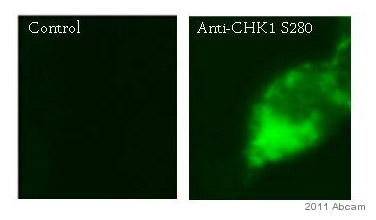
ab59988 staining phosphoChk1 (S280) in human colon adenocarcinoma cells (LoVo) by ICC/IF (Immunocytochemistry/immunofluorescence). Cells were fixed with paraformaldehyde, permeabilized with Triton 0.1% and blocked with 1% BSA for 2 hours at 20°C. Samples were incubated with primary antibody (1/250 in PBS + 0.1% Triton) for 3 hours at 20°C. A 1.4 nm Gold (ultra-small) FITC-conjugated Goat anti-rabbit polyclonal (1/500) was used as the secondary antibody.