![All lanes : Anti-CHD1L antibody [EPR14515(2)] (ab197019) at 1/5000 dilutionLane 1 : HeLa (Human epithelial cells from cervix adenocarcinoma) cell lysateLane 2 : A549 (Human lung carcinoma) cell lysateLysates/proteins at 20 µg per lane.SecondaryGoat Anti-Rabbit IgG, (H+L), Peroxidase conjugated at 1/1000 dilution](http://www.bioprodhub.com/system/product_images/ab_products/2/sub_1/28150_ab197019-236675-ab197019WB.jpg)
All lanes : Anti-CHD1L antibody [EPR14515(2)] (ab197019) at 1/5000 dilutionLane 1 : HeLa (Human epithelial cells from cervix adenocarcinoma) cell lysateLane 2 : A549 (Human lung carcinoma) cell lysateLysates/proteins at 20 µg per lane.SecondaryGoat Anti-Rabbit IgG, (H+L), Peroxidase conjugated at 1/1000 dilution
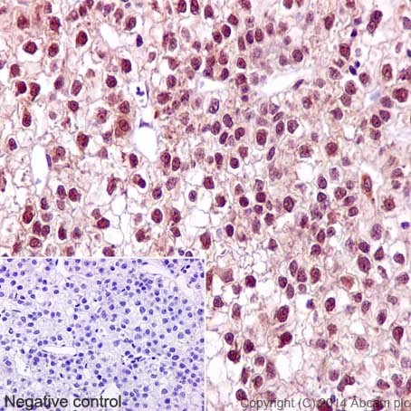
Immunohistochemical analysis of paraffin-embedded Human hepatocellular carcinoma tissue labeling CHD1L with ab197019 at 1/500 dilution, followed by Goat Anti-Rabbit IgG H&L (HRP) (ab97051) secondary antibody at 1/500 dilution. Nuclear and weakly cytoplasm staining on Human hepatocellular carcinoma tissue is observed. Counter stained with Hematoxylin.Negative control: Used PBS instead of primary antibody, secondary antibody is Goat Anti-Rabbit IgG H&L (HRP) (ab97051) at 1/500 dilution.
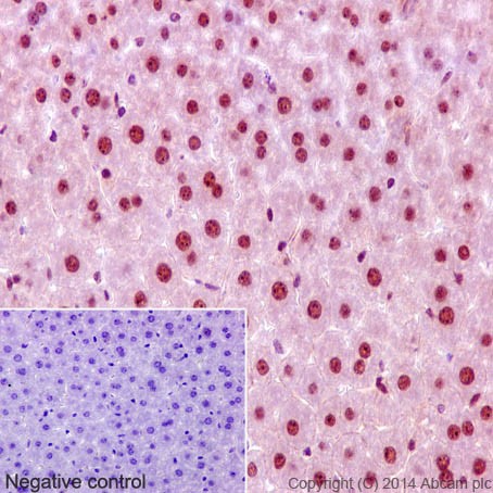
Immunohistochemical analysis of paraffin-embedded mouse liver tissue labeling CHD1L with ab197019 at 1/500 dilution, followed by Goat Anti-Rabbit IgG H&L (HRP) (ab97051) secondary antibody at 1/500 dilution. Nuclear and weakly cytoplasm staining on mouse liver tissue is observed. Counter stained with Hematoxylin.Negative control: Used PBS instead of primary antibody, secondary antibody is Goat Anti-Rabbit IgG H&L (HRP) (ab97051) at 1/500 dilution.
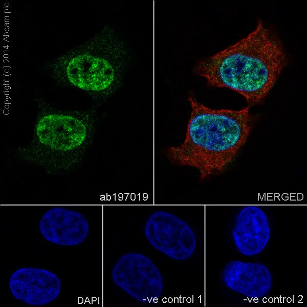
Immunofluorescent analysis of 4% paraformaldehyde-fixed, 0.1% Triton X-100 permeabilized HeLa (Human epithelial cells from cervix adenocarcinoma) cells labeling CHD1L with ab197019 at 1/500 dilution, followed by Goat anti-rabbit IgG (Alexa Fluor® 488) (ab150077) secondary antibody at 1/500 dilution (green). Nuclear and weakly cytoplasm staining on HeLa cell line was observed. The nuclear counter stain is DAPI (blue). Tubulin is detected with ab7291 (anti-Tubulin mouse mAb) at 1/1000 dilution and ab150120 (AlexaFluor®594 Goat anti-Mouse secondary) at 1/500 dilution (red).The negative controls are as follows:-ve control 1: ab197019 at 1/500 dilution followed by ab150120 (AlexaFluor®594 Goat anti-Mouse secondary) at 1/500 dilution.-ve control 2: ab7291 (anti-Tubulin mouse mAb) at 1/1000 dilution followed by ab150077 (Alexa Fluor®488 Goat Anti-Rabbit IgG H&L) at 1/500 dilution.
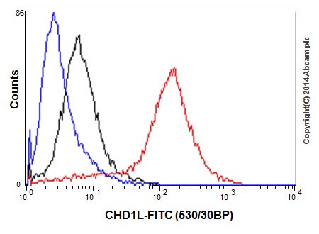
Flow cytometric analysis of HeLa (Human epithelial cells from cervix adenocarcinoma) cells labeling CHD1L with ab197019 at 1/520 dilution (red) compared with a rabbit monoclonal IgG isotype control (black) and an unlabelled control (cells without incubation with primary antibody and secondary antibody; blue). Goat anti rabbit IgG (FITC) at 1/150 dilution was used as the secondary antibody.
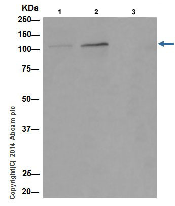
CHD1L was immunoprecipitated from 293 (Human embryonic kidney) whole cell extract with ab197019 at 1/30 dilution. Western blot was performed from the immunoprecipitate using ab197019 at 1/1000 dilution. Anti-Rabbit IgG (HRP), specific to the non-reduced form of IgG, was used as secondary antibody at 1/1500 dilution. Lane 1: 293 whole cell extract (Input) 10 µg. Lane 2: ab197019 IP in 293 whole cell extract. Lane 3: Rabbit monoclonal IgG (ab172730) instead of ab197019 in 293 whole cell extract.Blocking and dilution buffer and concentration: 5% NFDM/TBST.