![All lanes : Anti-Cdk1 + Cdk2 (phospho T14) antibody [EPR17499] (ab183550) at 1/1000 dilutionLane 1 : HeLa (Human epithelial cells from cervix adenocarcinoma) whole cell lysate treated with 2mg/ml hydroxyurea for 24 hours.Lane 2 : Untreated HeLa whole cell lysateLysates/proteins at 10 µg per lane.SecondaryGoat Anti-Rabbit IgG, (H+L),Peroxidase conjugated at 1/1000 dilution](http://www.bioprodhub.com/system/product_images/ab_products/2/sub_1/26600_ab183550-237990-183550WB.jpg)
All lanes : Anti-Cdk1 + Cdk2 (phospho T14) antibody [EPR17499] (ab183550) at 1/1000 dilutionLane 1 : HeLa (Human epithelial cells from cervix adenocarcinoma) whole cell lysate treated with 2mg/ml hydroxyurea for 24 hours.Lane 2 : Untreated HeLa whole cell lysateLysates/proteins at 10 µg per lane.SecondaryGoat Anti-Rabbit IgG, (H+L),Peroxidase conjugated at 1/1000 dilution
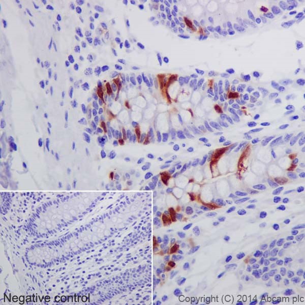
Immunohistochemical analysis of paraffin-embedded Human colon tissue labeling Cdk1 + Cdk2 (phospho T14) with ab183550 at 1/250 dilution, followed by Goat Anti-Rabbit IgG H&L (HRP) (ab97051) secondary antibody at 1/500 dilution. Scattered nuclear and cytoplasmic staining on epithelial cells of Human colon tissue is observed. Counter stained with Hematoxylin.Negative control: Used PBS instead of primary antibody, secondary antibody is Goat Anti-Rabbit IgG H&L (HRP) (ab97051) at 1/500 dilution.
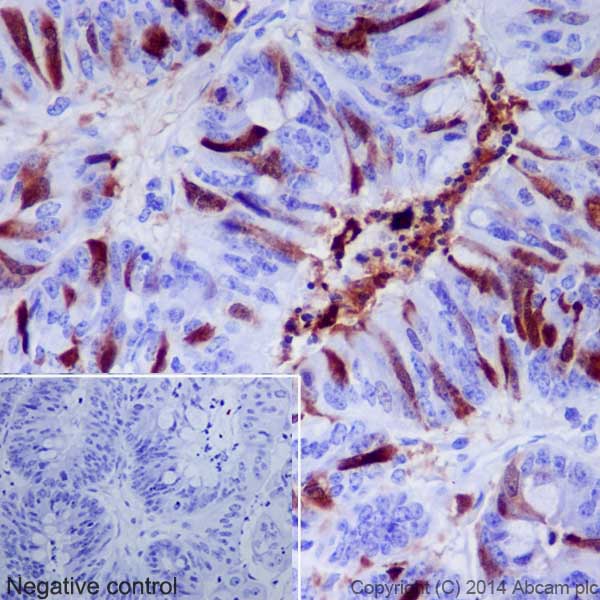
Immunohistochemical analysis of paraffin-embedded Human colonic adenocarcinoma tissue labeling Cdk1 + Cdk2 (phospho T14) with ab183550 at 1/250 dilution, followed by Goat Anti-Rabbit IgG H&L (HRP) (ab97051) secondary antibody at 1/500 dilution. Nuclear and cytoplasmic staining on cancer cells of Human colonic adenocarcinoma tissue is observed. Counter stained with Hematoxylin.Negative control: Used PBS instead of primary antibody, secondary antibody is Goat Anti-Rabbit IgG H&L (HRP) (ab97051) at 1/500 dilution.
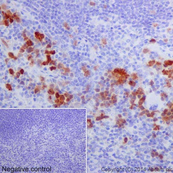
Immunohistochemical analysis of paraffin-embedded mouse spleen tissue labeling Cdk1 + Cdk2 (phospho T14) with ab183550 at 1/250 dilution, followed by Goat Anti-Rabbit IgG H&L (HRP) (ab97051) secondary antibody at 1/500 dilution. Nuclear and cytoplasmic staining on lymphocytes of mouse spleen tissue is observed. Counter stained with Hematoxylin.Negative control: Used PBS instead of primary antibody, secondary antibody is Goat Anti-Rabbit IgG H&L (HRP) (ab97051) at 1/500 dilution.
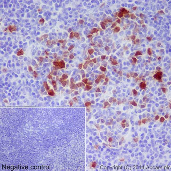
Immunohistochemical analysis of paraffin-embedded rat spleen tissue labeling Cdk1 + Cdk2 (phospho T14) with ab183550 at 1/250 dilution, followed by Goat Anti-Rabbit IgG H&L (HRP) (ab97051) secondary antibody at 1/500 dilution. Nuclear and cytoplasmic staining on lymphocytes of rat spleen tissue is observed. Counter stained with Hematoxylin.Negative control: Used PBS instead of primary antibody, secondary antibody is Goat Anti-Rabbit IgG H&L (HRP) (ab97051) at 1/500 dilution.
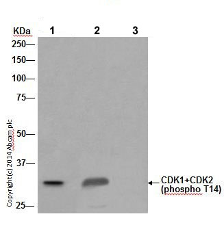
Cdk1 + Cdk2 (phospho T14) was immunoprecipitated from 1mg of HeLa (Human epithelial cells from cervix adenocarcinoma) whole cell extract with ab183550 at 1/40 dilution. Western blot was performed from the immunoprecipitate using ab183550 at 1/1000 dilution. Anti-Rabbit IgG (HRP), specific to the non-reduced form of IgG, was used as secondary antibody at 1/1000 dilution.Lane 1 (Input): HeLa whole cell extract 10 µg (Input).Lane 2: ab183550 IP in HeLa whole cell extract.Lane 3: Rabbit monoclonal IgG (ab172730) instead of ab183550 in HeLa whole cell extract.Blocking and dilution buffer and concentration: 5% NFDM/TBST.
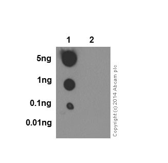
Dot blot analysis of Cdk1 + Cdk2 (phospho T14) peptide (Lane 1), and non-phospho peptide (Lane 2), labeled using ab183550 at 1/1000 dilution, followed by Goat Anti-Rabbit IgG, (H+L), Peroxidase conjugated secondary antibody at 1/1000 dilution.Blocking/Dilution buffer: 5% NFDM/TBST.Exposure time = 3 minutes