Anti-CD9 antibody [EPR2949]
| Name | Anti-CD9 antibody [EPR2949] |
|---|---|
| Supplier | Abcam |
| Catalog | ab92726 |
| Prices | $403.00 |
| Sizes | 100 µl |
| Host | Rabbit |
| Clonality | Monoclonal |
| Isotype | IgG |
| Clone | EPR2949 |
| Applications | FC WB IP IHC-P ICC/IF |
| Species Reactivities | Mouse, Rat, Human |
| Antigen | A synthetic peptide corresponding to residues within Human CD9 |
| Description | Rabbit Monoclonal |
| Gene | CD9 |
| Conjugate | Unconjugated |
| Supplier Page | Shop |
Product images
![All lanes : Anti-CD9 antibody [EPR2949] (ab92726) at 1/1000 dilutionLane 1 : HeLa cell lysateLane 2 : HuT-78 cell lysateLane 3 : U87-MG cell lysateLysates/proteins at 10 µg per lane.SecondaryHRP labelled goat anti-rabbit IgG at 1/2000 dilution](http://www.bioprodhub.com/system/product_images/ab_products/2/sub_1/25981_CD9-Primary-antibodies-ab92726-1.JPG) All lanes : Anti-CD9 antibody [EPR2949] (ab92726) at 1/1000 dilutionLane 1 : HeLa cell lysateLane 2 : HuT-78 cell lysateLane 3 : U87-MG cell lysateLysates/proteins at 10 µg per lane.SecondaryHRP labelled goat anti-rabbit IgG at 1/2000 dilution
All lanes : Anti-CD9 antibody [EPR2949] (ab92726) at 1/1000 dilutionLane 1 : HeLa cell lysateLane 2 : HuT-78 cell lysateLane 3 : U87-MG cell lysateLysates/proteins at 10 µg per lane.SecondaryHRP labelled goat anti-rabbit IgG at 1/2000 dilution
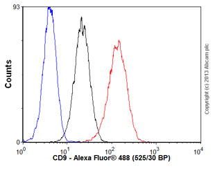 Overlay histogram showing Jurkat cells stained with ab92726 (red line). The cells were fixed with 80% methanol (5 min) and then permeabilized with 0.1% PBS-Tween for 20 min. The cells were then incubated in 1x PBS / 10% normal goat serum / 0.3M glycine to block non-specific protein-protein interactions followed by the antibody (ab92726, 1/1000 dilution) for 30 min at 22°C. The secondary antibody used was Alexa Fluor® 488 goat anti-rabbit IgG (H+L) (ab150077) at 1/2000 dilution for 30 min at 22°C. Isotype control antibody (black line) was rabbit IgG (monoclonal) (1μg/1x106 cells) used under the same conditions. Unlabelled sample (blue line) was also used as a control. Acquisition of >5,000 events were collected using a 20mW Argon ion laser (488nm) and 525/30 bandpass filter.
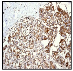 ab92726, at a 1/100 dilution, staining CD9 in paraffin embedded Human breast carcinoma tissue by Immunohistochemisty.
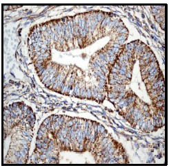 ab92726, at a 1/100 dilution, staining CD9 in paraffin embedded Human endometrial carcinoma tissue by Immunohistochemisty.
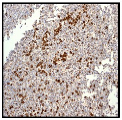 ab92726, at a 1/100 dilution, staining CD9 in paraffin embedded Human tonsil tissue by Immunohistochemisty.
 ab92726 showing positive staining in Papillary carcinoma of thyroid gland tissue.
 ab92726 showing positive staining in Normal kidney tissue.
 ab92726 showing positive staining in Astrocytoma tissue.
 ab92726 showing positive staining in Normal brain tissue.
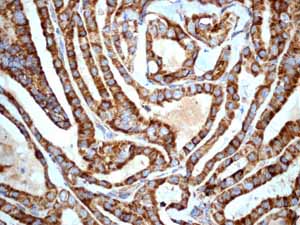
 ab92726 showing positive staining in Papillary carcinoma of thyroid gland tissue.
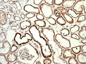 ab92726 showing positive staining in Normal kidney tissue.
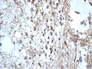
 ab92726 showing positive staining in Astrocytoma tissue.
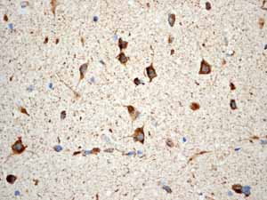 ab92726 showing positive staining in Normal brain tissue.
Product References
Identification of double-stranded genomic DNA spanning all chromosomes with - Identification of double-stranded genomic DNA spanning all chromosomes with
Kahlert C, Melo SA, Protopopov A, Tang J, Seth S, Koch M, Zhang J, Weitz J, Chin L, Futreal A, Kalluri R. J Biol Chem. 2014 Feb 14;289(7):3869-75.
Tumor-derived exosomes are enriched in DeltaNp73, which promotes oncogenic - Tumor-derived exosomes are enriched in DeltaNp73, which promotes oncogenic
Soldevilla B, Rodriguez M, San Millan C, Garcia V, Fernandez-Perianez R, Gil-Calderon B, Martin P, Garcia-Grande A, Silva J, Bonilla F, Dominguez G. Hum Mol Genet. 2014 Jan 15;23(2):467-78.
CD9 tetraspanin interacts with CD36 on the surface of macrophages: a possible - CD9 tetraspanin interacts with CD36 on the surface of macrophages: a possible
Huang W, Febbraio M, Silverstein RL. PLoS One. 2011;6(12):e29092.