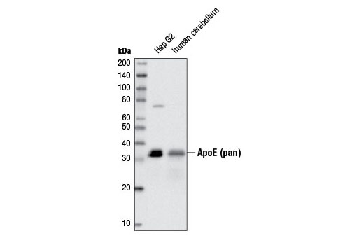
Western blot analysis of cell extracts from Hep G2 cells treated with Brefeldin A #9972 (10 ng/ml, 90 min) and human cerebellum using ApoE (pan) (D7I9N) Rabbit mAb.

Western blot analysis of cell extracts from 293T cells, treated with Brefeldin A #9972 (10 ng/ml, 90 min; +) and mock transfected (-) or transfected with a construct expressing full-length human ApoE2 (hApoE2; +), ApoE3 (hApoE3; +), or ApoE4 (hApoE4; +), using ApoE (pan) (D7I9N) Rabbit mAb (upper) and α-Actinin (D6F6) XP ® Rabbit mAb #6487 (lower).

Immunoprecipitation of ApoE from Hep G2 cells treated with Brefeldin A #9972 (10 ng/ml, 90 min), using Rabbit (DA1E) mAb IgG XP ® Isotype Control #3900 (lane 2) or ApoE (pan) (D7I9N) Rabbit mAb (lane 3). Lane 1 is 10% input. Western blot analysis was performed using ApoE (pan) (D7I9N) Rabbit mAb.

Immunohistochemical analysis of paraffin-embedded human small intestine using ApoE (pan) (D7I9N) Rabbit mAb.

Immunohistochemical analysis of paraffin-embedded human kidney using ApoE (pan) (D7I9N) Rabbit mAb.

Immunohistochemical analysis of paraffin-embedded human skin using ApoE (pan) (D7I9N) Rabbit mAb in the presence of control peptide (left) and antigen-specific peptide (right).

Immunohistochemical analysis of paraffin-embedded human spleen using ApoE (pan) (D7I9N) Rabbit mAb.