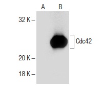
Cdc42 (B-8): sc-8401. Western blot analysis of Cdc42 expression in Jurkat (A) and 3611-RF (B) whole cell lysates.
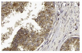
Cdc42 (B-8): sc-8401 Immunoperoxidase staining of formalin fixed, paraffin-embedded human prostate tissue showing cytoplasmic staining of glandular cells. Kindly provided by The Swedish Human Protein Atlas (HPA) program.
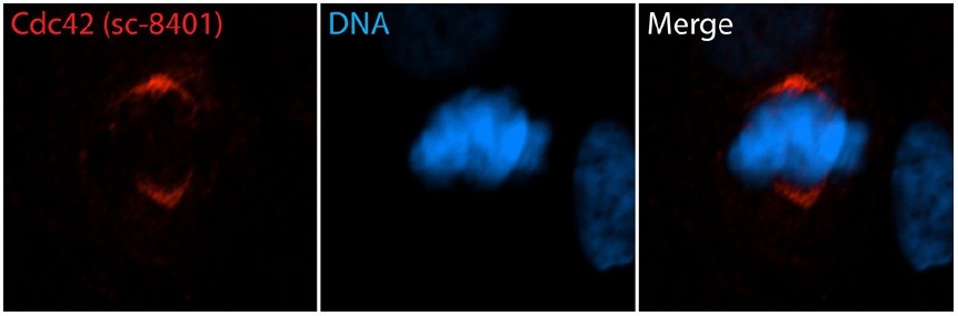
Cdc42 (B-8): sc-8401. Immunofluorescent staining of formalin fixed human retinal pigment epithelial (RPE) cells showing mitotic spindle staining (red immunofluorescence) and Hoechst 33342: sc-391054 nuclear counterstain (blue immunofluorescence). Secondary antibody used was donkey anti-mouse IgG-CFL 555: sc-362268 . Image kindly provided by Moshe Kim, Department of Cell Biology, Hospital for Sick Children (Toronto, Canada)
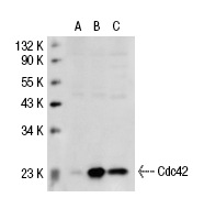
Cdc42 (B-8): sc-8401. Western blot analysis of Cdc42 expression in non-transfected 293T: sc-117752 (A), human Cdc42 transfected 293T: sc-110467 (B) and Jurkat (C) whole cell lysates.
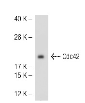
Cdc42 (B-8): sc-8401. Western blot analysis of Cdc42 expression in Jurkat whole cell lysate.
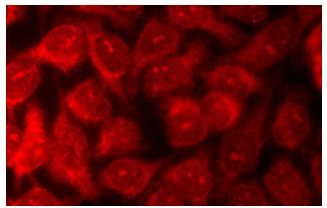
Cdc42 (B-8): sc-8401. Immunofluorescence staining of methanol-fixed HeLa cells showing cytoplasmic localization.
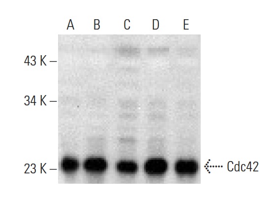
Cdc42 (B-8): sc-8401. Western blot analysis of Cdc42 expression in Jurkat (A), 3611-RF (B), HeLa (C), THP-1 (D) and A-431 (E) whole cell lysates.
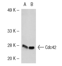
Cdc42 (B-8): sc-8401. Western blot analysis of Cdc42 expression in non-transfected: sc-117752 (A) and human Cdc42 transfected: sc-110467 (B) 293T whole cell lysates.
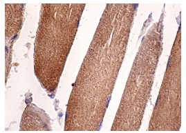
Cdc42 (B-8): sc-8401. Immunoperoxidase staining of formalin fixed, paraffin-embedded human skeletal muscle tissue showing cytoplasmic staining of myocytes.
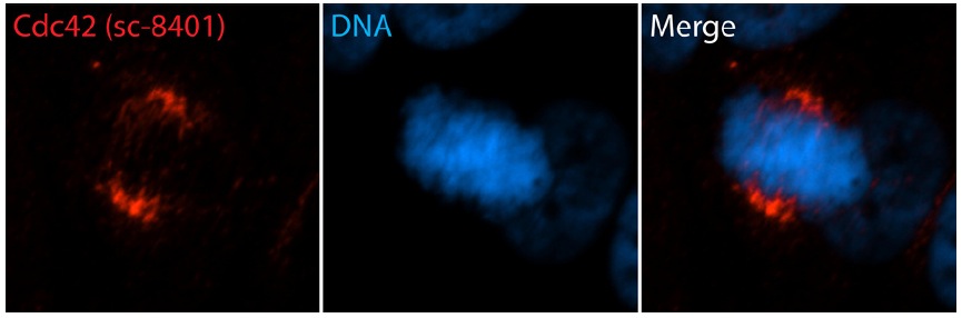
Cdc42 (B-8): sc-8401. Immunofluorescent staining of formalin fixed human retinal pigment epithelial (RPE) cells showing mitotic spindle staining (red immunofluorescence) and Hoechst 33342: sc-391054 nuclear counterstain (blue immunofluorescence). Secondary antibody used was donkey anti-mouse IgG-CFL 555: sc-362268 . Image kindly provided by Moshe Kim, Department of Cell Biology, Hospital for Sick Children (Toronto, Canada)