Anti-CD3 antibody
| Name | Anti-CD3 antibody |
|---|---|
| Supplier | Abcam |
| Catalog | ab16044 |
| Prices | $398.00 |
| Sizes | 100 µg |
| Host | Rabbit |
| Clonality | Polyclonal |
| Isotype | IgG |
| Applications | IP IHC-P IHC-F WB |
| Species Reactivities | Mouse, Rat, Human |
| Antigen | Synthetic peptide conjugated to KLH derived from within residues 150 to the C-terminus of Human CD3 |
| Description | Rabbit Polyclonal |
| Gene | CD3D |
| Conjugate | Unconjugated |
| Supplier Page | Shop |
Product images
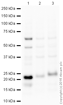 All lanes : Anti-CD3 antibody (ab16044) at 1 µg/mlLane 1 : Jurkat (Human T cell lymphoblast-like cell line) Whole Cell Lysate at 10 µgLane 2 : Thymus (Mouse) Tissue Lysate at 20 µgLane 3 : Thymus (Rat) Tissue Lysate at 20 µgSecondaryAnti-Rabbit IgG VHH Single Domain Antibody (HRP) (ab191866) at 1/50000 dilutiondeveloped using the ECL techniquePerformed under reducing conditions.
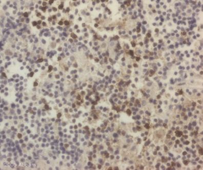 ab16044 staining CD3 in human thymus tissue section by Immunohistochemistry (Formalin/PFA-fixed paraffin-embedded sections). Tissue underwent fixation in formaldehyde, heat mediated antigen retrieval in buffer and blocking (5 minutes/peroxidase block then 10 minutes/protein block) for 45 minutes at 20°C.The primary antibody was diluted, 1/250 and incubated with sample for 45 minutes at 20°C. A HRP conjugated goat polyclonal to rabbit IgG was used undiluted as secondary.See Abreview
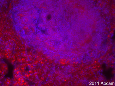 Rabbit polyclonal antibody (ab16044) detecting CD3 protein on PFA perfused Rat spleen section. The section was blocked with 10% donkey serum in PBS for 1hour at 24°C and then incubated with a 1/200 dilution of ab16044 for 24 hours at 4°C. Secondary antibody is a donkey anti-rabbit Alexa 568 (1/1000) and nuclei stained using the Hoechst method are depicted in blue . CD3 expressed in T cells but not in the B-cell germinal centre.See Abreview
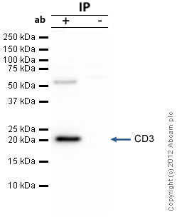 CD3 was immunoprecipitated using 0.5mg Jurkat whole cell extract, 5µg of Rabbit polyclonal to CD3 and 50µl of protein G magnetic beads (+). No antibody was added to the control (-). The antibody was incubated under agitation with Protein G beads for 10min, Jurkat whole cell extract lysate diluted in RIPA buffer was added to each sample and incubated for a further 10min under agitation.Proteins were eluted by addition of 40µl SDS loading buffer and incubated for 10min at 70oC; 10µl of each sample was separated on a SDS PAGE gel, transferred to a nitrocellulose membrane, blocked with 5% BSA and probed with ab16044.Secondary: Clean-Blot IP Detection Reagent (HRP) at 1/500 dilution.Band: 23kDa; CD3
Product References
Deficiency of the negative immune regulator B7-H1 enhances inflammation and - Deficiency of the negative immune regulator B7-H1 enhances inflammation and
Uceyler N, Gobel K, Meuth SG, Ortler S, Stoll G, Sommer C, Wiendl H, Kleinschnitz C. Exp Neurol. 2010 Mar;222(1):153-60.
CXC chemokine receptor 4 expressed in T cells plays an important role in the - CXC chemokine receptor 4 expressed in T cells plays an important role in the
Chung SH, Seki K, Choi BI, Kimura KB, Ito A, Fujikado N, Saijo S, Iwakura Y. Arthritis Res Ther. 2010;12(5):R188.
Macrophage colony stimulating factor is a crucial factor for the intrinsic - Macrophage colony stimulating factor is a crucial factor for the intrinsic
Muller M, Berghoff M, Kobsar I, Kiefer R, Martini R. Exp Neurol. 2007 Jan;203(1):55-62. Epub 2006 Sep 8.
Immune cells contribute to myelin degeneration and axonopathic changes in mice - Immune cells contribute to myelin degeneration and axonopathic changes in mice
Ip CW, Kroner A, Bendszus M, Leder C, Kobsar I, Fischer S, Wiendl H, Nave KA, Martini R. J Neurosci. 2006 Aug 2;26(31):8206-16.