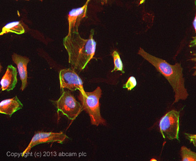
ICC/IF image of ab103404 stained HeLa cells. The cells were 100% methanol fixed (5 min) and then incubated in 1%BSA / 10% normal goat serum / 0.3M glycine in 0.1% PBS-Tween for 1h to permeabilise the cells and block non-specific protein-protein interactions. The cells were then incubated with the antibody ab103404 at 5µg/ml overnight at +4°C. The secondary antibody (green) was DyLight® 488 goat anti- rabbit (ab96899) IgG (H+L) used at a 1/250 dilution for 1h. Alexa Fluor® 594 WGA was used to label plasma membranes (red) at a 1/200 dilution for 1h. DAPI was used to stain the cell nuclei (blue) at a concentration of 1.43µM.
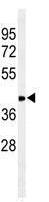
Anti-CCR7 antibody (ab103404) at 1/100 dilution + 293 cell line lysates at 35 µg
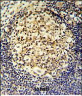
Immunohistochemical analysis of CCR7 in formalin fixed and paraffin embedded human tonsil tissue, using a 1/50 dilution of ab103404 followed by peroxidase conjugation of the secondary antibody and DAB staining.
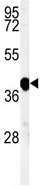
Anti-CCR7 antibody (ab103404) at 1/100 dilution + Mouse spleen tissue lysates at 35 µg