![Overlay histogram showing HeLa cells stained with ab125688 (red line). The cells were fixed with 80% methanol (5 min) and then permeabilized with 0.1% PBS-Tween for 20 min. The cells were then incubated in 1x PBS / 10% normal goat serum / 0.3M glycine to block non-specific protein-protein interactions followed by the antibody (ab125688, 1/100 dilution) for 30 min at 22°C. The secondary antibody used was Alexa Fluor® 488 goat anti-mouse IgG (H+L) (ab150113) at 1/2000 dilution for 30 min at 22°C. Isotype control antibody (black line) was mouse IgG1 [ICIGG1] (ab91353, 1μg/1x106 cells) used under the same conditions. Unlabelled sample (blue line) was also used as a control. Acquisition of >5,000 events were collected using a 20mW Argon ion laser (488nm) and 525/30 bandpass filter.](http://www.bioprodhub.com/system/product_images/ab_products/2/sub_1/21493_ab125688-12-ab125688FC.jpg)
Overlay histogram showing HeLa cells stained with ab125688 (red line). The cells were fixed with 80% methanol (5 min) and then permeabilized with 0.1% PBS-Tween for 20 min. The cells were then incubated in 1x PBS / 10% normal goat serum / 0.3M glycine to block non-specific protein-protein interactions followed by the antibody (ab125688, 1/100 dilution) for 30 min at 22°C. The secondary antibody used was Alexa Fluor® 488 goat anti-mouse IgG (H+L) (ab150113) at 1/2000 dilution for 30 min at 22°C. Isotype control antibody (black line) was mouse IgG1 [ICIGG1] (ab91353, 1μg/1x106 cells) used under the same conditions. Unlabelled sample (blue line) was also used as a control. Acquisition of >5,000 events were collected using a 20mW Argon ion laser (488nm) and 525/30 bandpass filter.
![All lanes : Anti-Catalase antibody [1B6] (ab125688) at 1/200 dilutionLane 1 : HEK293T cell lysate transfected with pCMV6-ENTRY control cDNALane 2 : HEK293T cell lysate transfected with pCMV6-ENTRY Catalase cDNALysates/proteins at 5 µg per lane.](http://www.bioprodhub.com/system/product_images/ab_products/2/sub_1/21494_Catalase-Primary-antibodies-ab125688-1.jpg)
All lanes : Anti-Catalase antibody [1B6] (ab125688) at 1/200 dilutionLane 1 : HEK293T cell lysate transfected with pCMV6-ENTRY control cDNALane 2 : HEK293T cell lysate transfected with pCMV6-ENTRY Catalase cDNALysates/proteins at 5 µg per lane.
![All lanes : Anti-Catalase antibody [1B6] (ab125688) at 1/200 dilutionLane 1 : HepG2 cell extractLane 2 : HeLa cell extractLane 3 : HT29 cell extractLane 4 : A549 cell extractLane 5 : COS7 cell extractLane 6 : Jurkat cell extractLane 7 : MDCK cell extractLane 8 : PC12 cell extractLane 9 : MCF7 cell extractLysates/proteins at 35 µg per lane.](http://www.bioprodhub.com/system/product_images/ab_products/2/sub_1/21495_Catalase-Primary-antibodies-ab125688-2.jpg)
All lanes : Anti-Catalase antibody [1B6] (ab125688) at 1/200 dilutionLane 1 : HepG2 cell extractLane 2 : HeLa cell extractLane 3 : HT29 cell extractLane 4 : A549 cell extractLane 5 : COS7 cell extractLane 6 : Jurkat cell extractLane 7 : MDCK cell extractLane 8 : PC12 cell extractLane 9 : MCF7 cell extractLysates/proteins at 35 µg per lane.
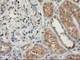
ab125688, at a dilution of 1/150, staining Catalase in paraffin-embedded Human kidney carcinoma tissue by Immunohistochemistry.
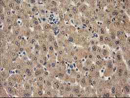
ab125688, at a dilution of 1/150, staining Catalase in paraffin-embedded Human liver tissue by Immunohistochemistry.
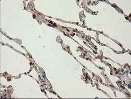
ab125688, at a dilution of 1/150, staining Catalase in paraffin-embedded Human lung tissue by Immunohistochemistry.
ab125688, at a dilution of 1/150, staining Catalase in paraffin-embedded Human endometrium tissue by Immunohistochemistry.
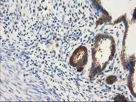
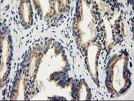
ab125688, at a dilution of 1/150, staining Catalase in paraffin-embedded Human prostate tissue by Immunohistochemistry.
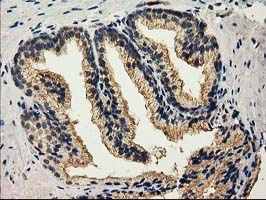
ab125688, at a dilution of 1/150, staining Catalase in paraffin-embedded Human prostate carcinoma tissue by Immunohistochemistry.
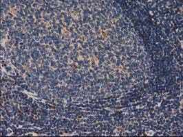
ab125688, at a dilution of 1/150, staining Catalase in paraffin-embedded Human lymph node by Immunohistochemistry.
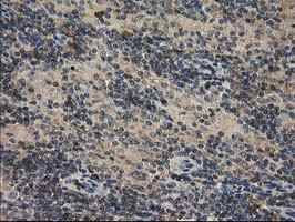
ab125688, at a dilution of 1/150, staining Catalase in paraffin-embedded Human lymphoma tissue by Immunohistochemistry.
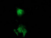
ab125688, at 1/100 dilution, staining Catalase in COS7 cells transiently transfected by pCMV6-ENTRY Catalase by Immunofluorescence.