![All lanes : Anti-Caspase-14 antibody [EPR12927] (ab174847) at 1/1000 dilutionLane 1 : MCF7 cell lysateLane 2 : ECV-304 cell lysateLane 3 : HACAT cell lysateLane 4 : Human skin lysateLysates/proteins at 10 µg per lane.](http://www.bioprodhub.com/system/product_images/ab_products/2/sub_1/21143_ab174847-200925-ab1748471.JPG)
All lanes : Anti-Caspase-14 antibody [EPR12927] (ab174847) at 1/1000 dilutionLane 1 : MCF7 cell lysateLane 2 : ECV-304 cell lysateLane 3 : HACAT cell lysateLane 4 : Human skin lysateLysates/proteins at 10 µg per lane.
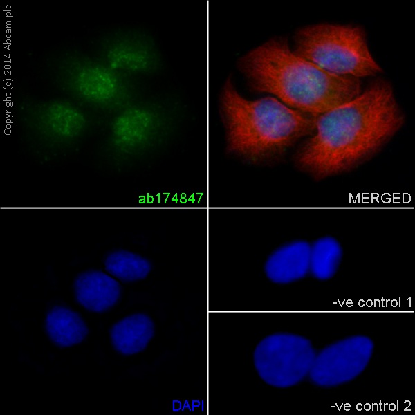
Immunofluorescent staining of HaCaT cells labeling Caspase-14 (green) with ab174847 at 1:250 dilution, and labeling tubulin (red) with ab7291 at 1:500 (2 µg/ml). Goat Anti-Rabbit IgG H&L (Alexa Fluor® 488) (ab150077) was used at 1:200 and Goat Anti-Mouse IgG H&L (Alexa Fluor® 594) (ab150120) was used at 1:200.-ve control 1– Rabbit primary and anti-mouse secondary antibody. -ve control 2 – Mouse primary antibody and anti-rabbit secondary antibody.
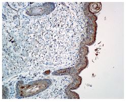
Immunohistochemical analysis of paraffin-embedded fetal skin tissue labeling Caspase-14 using ab174847 at a 1:100 dilution.
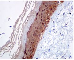
Immunohistochemical analysis of paraffin-embedded Human skin tissue labeling Caspase-14 using ab174847 at a 1:100 dilution.
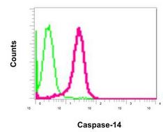
Flow cytometric analysis of permeabilized HACAT cells labeling Caspase-14 using ab174847 at a 1:10 dilution (red), or rabbit IgG negative control (green).
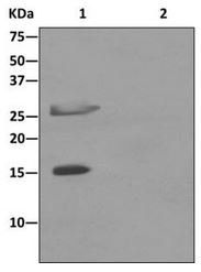
Western blot analysis on immunoprecipitation pellet from fetal skin lysate (lane 1) or 1X PBS (lane 2, negative control) using ab174847 at 1:10 dilution, and HRP-conjugated anti-rabbit IgG preferentially detecting the non-reduced form of rabbit IgG.