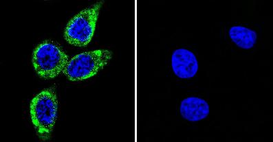
Immunofluorescence analysis of U251 cells labeling phosphorylated CaMKII (green) with ab171095 at a 1/100 dilution. Nuclei are stained (blue).
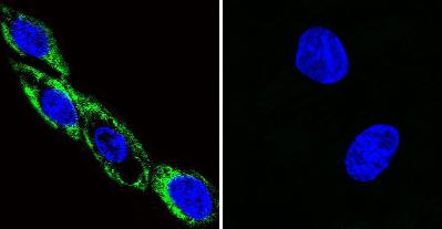
Immunofluorescence analysis of HeLa cells labeling phosphorylated CaMKII (green) with ab171095 at a 1/50 dilution. Nuclei are stained (blue).
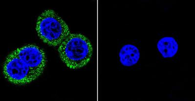
Immunofluorescence analysis of C6 cells labeling phospohrylated CaMKII (green) with ab171095 at a 1/100 dilution. Nuclei are stained (blue).
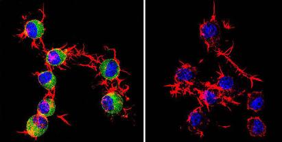
Immunofluorescence analysis of Neuro-2 cells (left image) or cells stimulated with Lambda-phosphatase (right image) labeling phosphorylated CaMKII (green) with ab171095 at a 1/50 dilution. F-actin (red) and nuclei (blue).
![Lanes 1 - 2 : Anti-CaMKII (phospho T286) antibody [22B1] (ab171095) at 1/2000 dilutionLanes 3 - 4 : A non phosphorylated CaMKII antibody at 1/2000 dilutionLane 1 : Unphosphorylated rat brain homogenateLane 2 : Phosphorylated Rat brain homogenateLane 3 : Unphosphorylated Rat brain homogenateLane 4 : Phosphorylated Rat brain homogenate](http://www.bioprodhub.com/system/product_images/ab_products/2/sub_1/20002_ab171095-171597-ab171095wb1.jpg)
Lanes 1 - 2 : Anti-CaMKII (phospho T286) antibody [22B1] (ab171095) at 1/2000 dilutionLanes 3 - 4 : A non phosphorylated CaMKII antibody at 1/2000 dilutionLane 1 : Unphosphorylated rat brain homogenateLane 2 : Phosphorylated Rat brain homogenateLane 3 : Unphosphorylated Rat brain homogenateLane 4 : Phosphorylated Rat brain homogenate