Anti-Calcium Pump PMCA2 ATPase antibody
| Name | Anti-Calcium Pump PMCA2 ATPase antibody |
|---|---|
| Supplier | Abcam |
| Catalog | ab3529 |
| Prices | $401.00 |
| Sizes | 100 µg |
| Host | Rabbit |
| Clonality | Polyclonal |
| Isotype | IgG |
| Applications | ICC/IF ICC/IF IHC-P IHC-F ICC/IF IP WB ELISA IHC-F |
| Species Reactivities | Mouse, Rat, Chicken, Human, Pig, Primate |
| Antigen | Synthetic peptide corresponding to Human Calcium Pump PMCA2 ATPase aa 5-19 |
| Description | Rabbit Polyclonal |
| Gene | ATP2B2 |
| Conjugate | Unconjugated |
| Supplier Page | Shop |
Product images
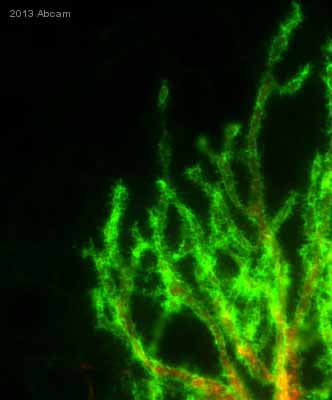 ab3529 staining Calcium Pump PMCA2 ATPase in Mouse purkinje cells in cerebellum tissue sections by Immunohistochemistry (IHC-P - paraformaldehyde-fixed, paraffin-embedded sections). Tissue was fixed with paraformaldehyde and permeabilized with 0.5% Triton X-100. Samples were incubated with primary antibody (1/500 in PBS + 3% NGS + 0.5% Triton X-100) for 16 hours at 4°C. An Alexa Fluor® 488-conjugated Goat anti-rabbit IgG monoclonal (1/500) was used as the secondary antibody.See Abreview
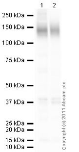 All lanes : Anti-Calcium Pump PMCA2 ATPase antibody (ab3529) at 1 µg/mlLane 1 : Brain (Rat) Tissue LysateLane 2 : Brain (Human) Tissue Lysate - adult normal tissue (ab29466)Lysates/proteins at 10 µg per lane.SecondaryGoat Anti-Rabbit IgG H&L (HRP) preadsorbed (ab97080) at 1/5000 dilutiondeveloped using the ECL techniquePerformed under reducing conditions.
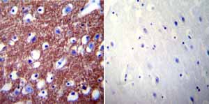 Immunohistochemistry was performed on normal biopsies of deparaffinized Human brain tissue. To expose target proteins heat induced antigen retrieval was performed using 10mM sodium citrate (pH6.0) buffer microwaved for 8-15 minutes. Following antigen retrieval tissues were blocked in 3% BSA-PBS for 30 minutes at room temperature. Tissues were then probed at a dilution of 1:200 with a rabbit polyclonal antibody recognizing PMCA2 ATPase ab3529 or without primary antibody (negative control) overnight at 4°C in a humidified chamber. Tissues were washed extensively with PBST and endogenous peroxidase activity was quenched with a peroxidase suppressor. Detection was performed using a biotin-conjugated secondary antibody and SA-HRP followed by colorimetric detection using DAB. Tissues were counterstained with hematoxylin and prepped for mounting.
 Immunohistochemistry was performed on cancer biopsies of deparaffinized Human cervical carcinoma tissue. To expose target proteins heat induced antigen retrieval was performed using 10mM sodium citrate (pH6.0) buffer microwaved for 8-15 minutes. Following antigen retrieval tissues were blocked in 3% BSA-PBS for 30 minutes at room temperature. Tissues were then probed at a dilution of 1:20 with a rabbit polyclonal antibody recognizing PMCA2 ATPase ab3529 or without primary antibody (negative control) overnight at 4°C in a humidified chamber. Tissues were washed extensively with PBST and endogenous peroxidase activity was quenched with a peroxidase suppressor. Detection was performed using a biotin-conjugated secondary antibody and SA-HRP followed by colorimetric detection using DAB. Tissues were counterstained with hematoxylin and prepped for mounting.
Product References
The plasma membrane Ca2+-ATPase2 (PMCA2) is involved in the regulation of - The plasma membrane Ca2+-ATPase2 (PMCA2) is involved in the regulation of
Sherkhane P, Kapfhammer JP. Neural Plast. 2013;2013:321685.
Reduced expression of plasma membrane calcium ATPase 2 and collapsin response - Reduced expression of plasma membrane calcium ATPase 2 and collapsin response
Kurnellas MP, Li H, Jain MR, Giraud SN, Nicot AB, Ratnayake A, Heary RF, Elkabes S. Cell Death Differ. 2010 Sep;17(9):1501-10.
Presynaptic plasma membrane Ca2+ ATPase isoform 2a regulates excitatory synaptic - Presynaptic plasma membrane Ca2+ ATPase isoform 2a regulates excitatory synaptic
Jensen TP, Filoteo AG, Knopfel T, Empson RM. J Physiol. 2007 Feb 15;579(Pt 1):85-99. Epub 2006 Dec 14.