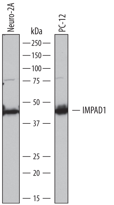
Detection of Mouse and Rat Inositol Monophosphatase 3/IMPAD1 by Western Blot. Western blot shows lysates of Neuro‑2A mouse neuroblastoma cell line and PC‑12 rat adrenal pheochromocytoma cell line. PVDF membrane was probed with 1 µg/mL of Sheep Anti-Mouse Inositol Monophosphatase 3/IMPAD1 Antigen Affinity-purified Polyclonal Antibody (Catalog # AF7028) followed by HRP-conjugated Anti-Sheep IgG Secondary Antibody (Catalog # HAF016). A specific band was detected for Inositol Monophosphatase 3/IMPAD1 at approximately 42 kDa (as indicated). This experiment was conducted under reducing conditions and using Immunoblot Buffer Group 1.
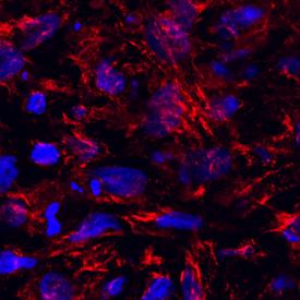
Inositol Monophosphatase 3/IMPAD1 in Mouse Mesenchymal Stem Cells. Inositol Monophosphatase 3/IMPAD1 was detected in immersion fixed mouse mesenchymal stem cells differentiated into chondrocytes using Sheep Anti-Mouse/Rat Inositol Monophosphatase 3/IMPAD1 Antigen Affinity-purified Polyclonal Antibody (Catalog # AF7028) at 10 µg/mL for 3 hours at room temperature. Cells were stained using the NorthernLights™ 557-conjugated Anti-Sheep IgG Secondary Antibody (red; Catalog # NL010) and counterstained with DAPI (blue). Specific staining was localized to cytoplasm. View our protocol for Fluorescent ICC Staining of Cells on Coverslips.