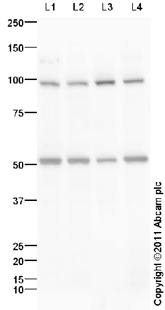
All lanes : Anti-Cadherin 8 antibody (ab97268) at 1 µg/mlLane 1 : SHSY-5Y (Human neuroblastoma cell line) Whole Cell LysateLane 2 : WERI (Human Retinoblastoma) Whole Cell LysateLane 3 : U-87 MG (Human glioblastoma astrocytoma) Whole Cell Lysate Lane 4 : SK N BE (Human neuroblastoma) Whole Cell Lysate Lysates/proteins at 10 µg per lane.SecondaryGoat Anti-Rabbit IgG H&L (HRP) preadsorbed (ab97080) at 1/5000 dilutiondeveloped using the ECL techniquePerformed under reducing conditions.
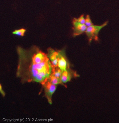
ICC/IF image of ab97268 stained U87MG cells. The cells were 4% formaldehyde fixed (10 min) and then incubated in 1%BSA / 10% normal goat serum / 0.3M glycine in 0.1% PBS-Tween for 1h to permeabilise the cells and block non-specific protein-protein interactions. The cells were then incubated with the antibody (ab97268, 5µg/ml) overnight at +4°C. The secondary antibody (green) was ab96899, DyLight® 488 goat anti-rabbit IgG (H+L) used at a 1/250 dilution for 1h. Alexa Fluor® 594 WGA was used to label plasma membranes (red) at a 1/200 dilution for 1h. DAPI was used to stain the cell nuclei (blue) at a concentration of 1.43µM.