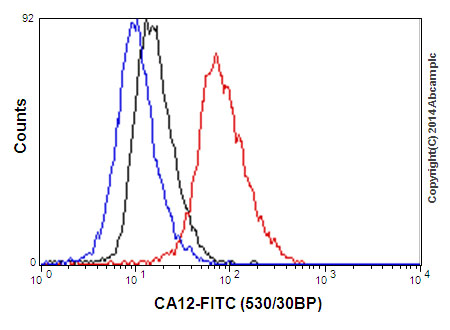
Flow cytometric analysis of SK-OV-3 cells (paraformaldehyde-fixed, 2%) labeling CA12 with ab195233 at 1/130 dilution (red) or a Rabbit monoclonal IgG (negative) (black), followed by Goat anti rabbit IgG (FITC) secondary at 1/150 dilution. Unlabeled cells (Blue).
Immunohistochemical analysis of paraffin-embedded Human stomach tissue labeling CA12 with ab195233 at 1/100 dilution followed by Goat Anti-Rabbit IgG H&L (HRP) (ab97051) at 1/500 dilution and counter-stained with Hematoxylin. (inset: negative control).Note: Cell membrane and cytoplasm staining on human stomach tissue was observed.
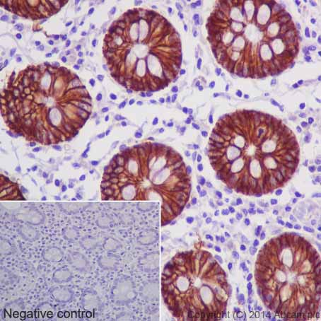
Immunohistochemical analysis of paraffin-embedded Human colon tissue labeling CA12 with ab195233 at 1/100 dilution followed by Goat Anti-Rabbit IgG H&L (HRP) (ab97051) at 1/500 dilution and counter-stained with Hematoxylin. (inset: negative control).Note: Cell membrane and cytoplasm staining on human colon tissue was observed.
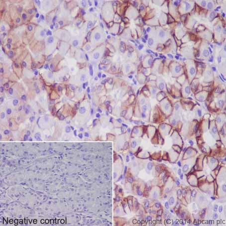
Immunohistochemical analysis of paraffin-embedded Human pancreas tissue labeling CA12 with ab195233 at 1/100 dilution followed by Goat Anti-Rabbit IgG H&L (HRP) (ab97051) at 1/500 dilution and counter-stained with Hematoxylin. (inset: negative control).Note: Cell membrane and weakly cytoplasm staining on human pancreas tissue was observed.
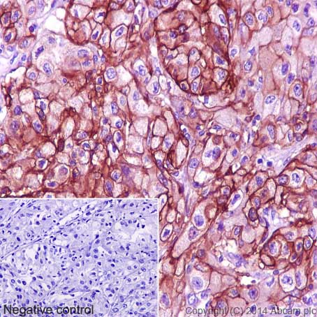
Immunohistochemical analysis of paraffin-embedded Human bladder transitional cell carcinoma tissue labeling CA12 with ab195233 at 1/100 dilution followed by Goat Anti-Rabbit IgG H&L (HRP) (ab97051) at 1/500 dilution and counter-stained with Hematoxylin. (inset: negative control).Note: Cell membrane and weakly cytoplasm staining on human transitional cell carcinoma of bladder tissue was observed.
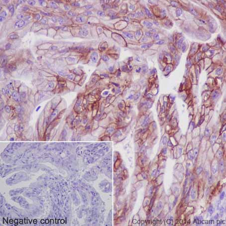
Immunohistochemical analysis of paraffin-embedded Human ovarian carcinoma tissue labeling CA12 with ab195233 at 1/100 dilution followed by Goat Anti-Rabbit IgG H&L (HRP) (ab97051) at 1/500 dilution and counter-stained with Hematoxylin. (inset: negative control).Note: Cell membrane and weakly cytoplasm staining on human ovarian carcinoma tissue was observed.
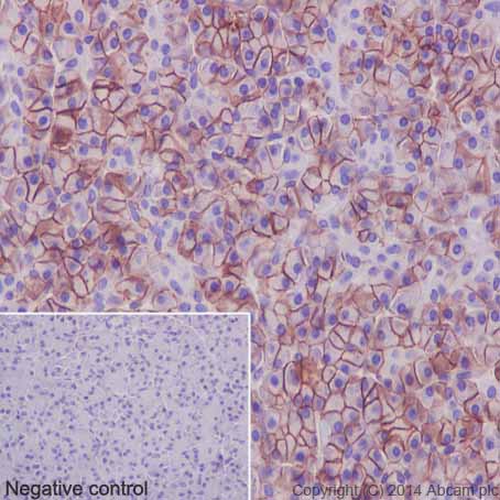
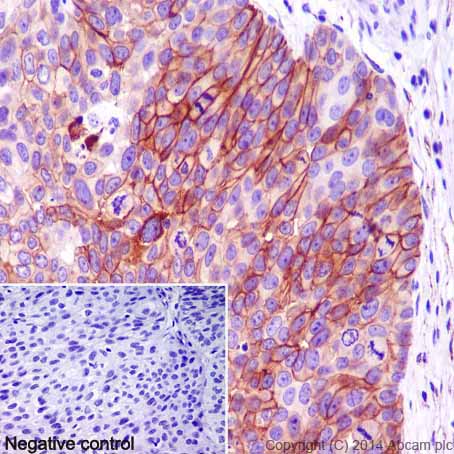
Immunohistochemical analysis of paraffin-embedded Human renal adenocarcinoma tissue labeling CA12 with ab195233 at 1/100 dilution followed by Goat Anti-Rabbit IgG H&L (HRP) (ab97051) at 1/500 dilution and counter-stained with Hematoxylin. (inset: negative control).Note: Cell membrane and cytoplasm staining on human renal adenocarcinoma tissue was observed.
![All lanes : Anti-CA12 antibody [EPR14861] - C-terminal (ab195233) at 1/10000 dilutionLane 1 : MCF7 cell lysateLane 2 : SKOV3 cell lysateLane 3 : 293 cell lysateLane 4 : HeLa cell lysateLane 5 : SKOV3 cell lysateLane 6 : Human fetal kidney lysateLane 7 : Human ovary cancer lysateLane 8 : Human breast cancer lysateLysates/proteins at 10 µg per lane.SecondaryGoat Anti-Rabbit IgG, (H+L), Peroxidase conjugate at 1/1000 dilutiondeveloped using the ECL technique](http://www.bioprodhub.com/system/product_images/ab_products/2/sub_1/18873_ab195233-234696-ab195233b.jpg)
All lanes : Anti-CA12 antibody [EPR14861] - C-terminal (ab195233) at 1/10000 dilutionLane 1 : MCF7 cell lysateLane 2 : SKOV3 cell lysateLane 3 : 293 cell lysateLane 4 : HeLa cell lysateLane 5 : SKOV3 cell lysateLane 6 : Human fetal kidney lysateLane 7 : Human ovary cancer lysateLane 8 : Human breast cancer lysateLysates/proteins at 10 µg per lane.SecondaryGoat Anti-Rabbit IgG, (H+L), Peroxidase conjugate at 1/1000 dilutiondeveloped using the ECL technique