![All lanes : Anti-C4 binding protein antibody [EPR17101] (ab199430) at 1/1000 dilutionLane 1 : Human plasmaLane 2 : Human ovary cancer extract.Lysates/proteins at 20 µg per lane.SecondaryAnti-Rabbit IgG (HRP), specific to the non-reduced form of IgG at 1/1000 dilution](http://www.bioprodhub.com/system/product_images/ab_products/2/sub_1/18172_ab199430-241313-199430.JPG)
All lanes : Anti-C4 binding protein antibody [EPR17101] (ab199430) at 1/1000 dilutionLane 1 : Human plasmaLane 2 : Human ovary cancer extract.Lysates/proteins at 20 µg per lane.SecondaryAnti-Rabbit IgG (HRP), specific to the non-reduced form of IgG at 1/1000 dilution
![Anti-C4 binding protein antibody [EPR17101] (ab199430) at 1/10000 dilution + Human serum at 20 µgSecondaryAnti-Rabbit IgG (HRP), specific to the non-reduced form of IgG at 1/1000 dilution](http://www.bioprodhub.com/system/product_images/ab_products/2/sub_1/18173_ab199430-241312-1994302.JPG)
Anti-C4 binding protein antibody [EPR17101] (ab199430) at 1/10000 dilution + Human serum at 20 µgSecondaryAnti-Rabbit IgG (HRP), specific to the non-reduced form of IgG at 1/1000 dilution
![Anti-C4 binding protein antibody [EPR17101] (ab199430) at 1/1000 dilution + Rat plasma at 10 µgSecondaryGoat Anti-Rabbit IgG, (H+L), Peroxidase conjugated at 1/1000 dilution](http://www.bioprodhub.com/system/product_images/ab_products/2/sub_1/18174_ab199430-241311-1994303.JPG)
Anti-C4 binding protein antibody [EPR17101] (ab199430) at 1/1000 dilution + Rat plasma at 10 µgSecondaryGoat Anti-Rabbit IgG, (H+L), Peroxidase conjugated at 1/1000 dilution
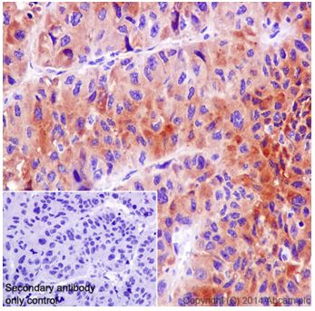
Immunohistochemical analysis of paraffin-embedded Human hepatocellular carcinoma tissue labeling C4 binding protein with ab199430 at 1/1000 dilution followed by Goat Anti-Rabbit IgG H&L (HRP) (ab97051) at 1/500 dilution. Cytoplasm staining on Human hepatocellular carcinoma tissue is observed. Counter stained with Hematoxylin.Negative control: Used PBS instead of primary ab, secondary ab is Goat Anti-Rabbit IgG H&L (HRP) (ab97051).
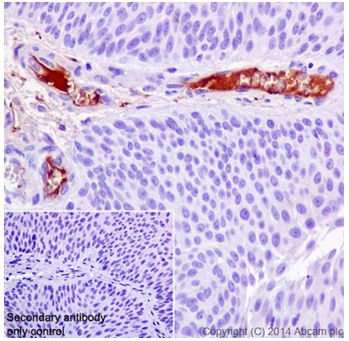
Immunohistochemical analysis of paraffin-embedded Human bladder carcinoma tissue labeling C4 binding protein with ab199430 at 1/1000 dilution followed by Goat Anti-Rabbit IgG H&L (HRP) (ab97051) at 1/500 dilution. Plasma staining on Human bladder carcinoma tissue is observed. Counter stained with Hematoxylin.Negative control: Used PBS instead of primary ab, secondary ab is Goat Anti-Rabbit IgG H&L (HRP) (ab97051).

Immunohistochemical analysis of paraffin-embedded Rat liver tissue labeling C4 binding protein with ab199430 at 1/1000 dilution followed by Goat Anti-Rabbit IgG H&L (HRP) (ab97051) at 1/500 dilution. Cytoplasm staining on rat liver tissue is observed. Counter stained with Hematoxylin.Negative control: Used PBS instead of primary ab, secondary ab is Goat Anti-Rabbit IgG H&L (HRP) (ab97051).
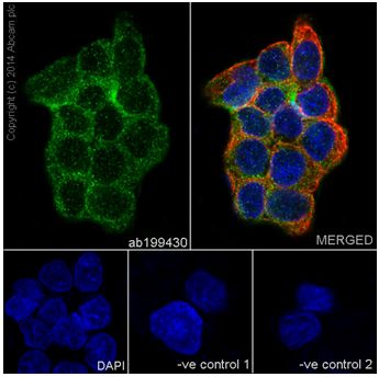
Immunofluorescent analysis of 4% paraformaldehyde-fixed, 0.1% Triton X-100 permeabilized HepG2 (Human liver hepatocellular carcinoma) cells labeling C4 binding protein with ab199430 at 1/250 dilution, followed by Goat anti-rabbit IgG (Alexa Fluor® 488) (ab150077) secondary antibody at 1/500 dilution (green). Confocal image showing cytoplasm staining on HepG2 cell line. The nuclear counterstain is DAPI (blue). Tubulin is detected with ab7291 (anti-Tubulin mouse mAb) at 1/1000 dilution and ab150120 (AlexaFluor®594 Goat anti-Mouse secondary) at 1/500 dilution (red).The negative controls are as follows:--ve control 1 - ab199430 at 1/250 dilution followed by ab150120 (AlexaFluor®594 Goat anti-Mouse secondary) at 1/500 dilution.-ve control 2. - ab7291 (anti-Tubulin mouse mAb) at 1/1000 dilution followed by ab150077 (Alexa Fluor®488 Goat Anti-Rabbit IgG H&L) at 1/500 dilution.
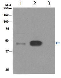
C4 binding protein was immunoprecipitated from 1mg of Human serum with ab199430 at 1/80 dilution. Western blot was performed from the immunoprecipitate using ab199430 at 1/10000 dilution. Anti-Rabbit IgG (HRP), specific to the non-reduced form of IgG, was used as secondary antibody at 1/1500 dilution. Lane 1: Human serum 10 µg. Lane 2: Human serum following immunoprecipitation. Lane 3: Rabbit monoclonal IgG (ab172730) instead of ab199430 in Human serum.Blocking and dilution buffer and concentration: 5% NFDM/TBST. Exposure: 10 seconds.