![All lanes : Anti-C1s antibody [4E3] (ab119759) at 1/2000 dilutionLane 1 : HEK293T cells were transfected with pCMV6-ENTRY control Lane 2 : HEK293T cells were transfected with pCMV6-ENTRY C1s cDNALysates/proteins at 5 µg per lane.](http://www.bioprodhub.com/system/product_images/ab_products/2/sub_1/17733_C1s-Primary-antibodies-ab119759-1.jpg)
All lanes : Anti-C1s antibody [4E3] (ab119759) at 1/2000 dilutionLane 1 : HEK293T cells were transfected with pCMV6-ENTRY control Lane 2 : HEK293T cells were transfected with pCMV6-ENTRY C1s cDNALysates/proteins at 5 µg per lane.
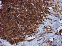
ab119759 at 1/150 dilution, staining C1s in formalin-fixed, paraffin-embedded adenocarcinoma of Human breast tissue by Immunohistochemistry.
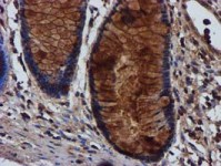
ab119759 at 1/150 dilution, staining C1s in formalin-fixed, paraffin-embedded Human colon tissue by Immunohistochemistry.
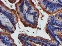
ab119759 at 1/150 dilution, staining C1s in formalin-fixed, paraffin-embedded adenocarcinoma of Human colon tissue by Immunohistochemistry.
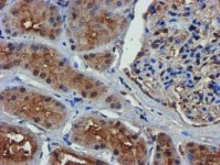
ab119759 at 1/150 dilution, staining C1s in formalin-fixed, paraffin-embedded Human kidney tissue by Immunohistochemistry.
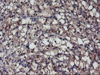
ab119759 at 1/150 dilution, staining C1s in formalin-fixed, paraffin-embedded carcinoma of Human kidney tissue by Immunohistochemistry.
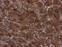
ab119759 at 1/150 dilution, staining C1s in formalin-fixed, paraffin-embedded carcinoma of Human liver tissue by Immunohistochemistry.
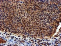
ab119759 at 1/150 dilution, staining C1s in formalin-fixed, paraffin-embedded carcinoma of Human bladder tissue by Immunohistochemistry.
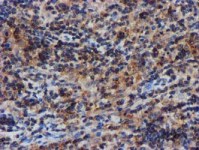
ab119759 at 1/150 dilution, staining C1s in formalin-fixed, paraffin-embedded Human lymphoma tissue by Immunohistochemistry.
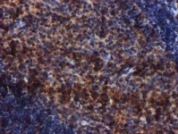
ab119759 at 1/150 dilution, staining C1s in formalin-fixed, paraffin-embedded Human tonsil tissue by Immunohistochemistry.
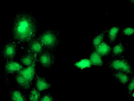
ab119759 at 1/100 dilution, staining C1s in COS7 cells transiently transfected by pCMV6-ENTRY C1s by Immunofluorescence.
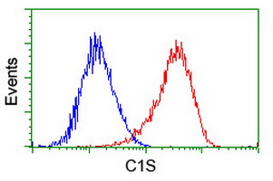
Flow cytometric Analysis of HeLa cells, using ab119759 at 1/100 dilution (Red), compared to a nonspecific negative control antibody (Blue).
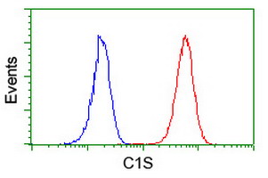
Flow cytometric Analysis of Jurkat cells, using ab119759 at 1/100 dilution (Red), compared to a nonspecific negative control antibody (Blue).