Human/Mouse Myeloperoxidase Affinity Purified Polyclonal Ab, Goat IgG
| Name | Human/Mouse Myeloperoxidase Affinity Purified Polyclonal Ab, Goat IgG |
|---|---|
| Supplier | R&D Systems |
| Catalog | AF3667-SP |
| Host | Goat |
| Clonality | Polyclonal |
| Isotype | IgG |
| Applications | IHC ICC/IF Simple Western WB |
| Species Reactivities | Human, Mouse |
| Antigen | Mouse myeloma cell line NS0-derived recombinant mouse MPO . Met16-Thr718 Accession Number P11247 |
| Description | Goat Polyclonal |
| Gene | MPO |
| Conjugate | Unconjugated |
| Supplier Page | Shop |
Product images
 Detection of Human/Mouse Myeloperoxidase/MPO by Western Blot. Western blot shows lysates of HL‑60 human acute promyelocytic leukemia cell line, human neutrophil cells, and mouse spleen tissue. PVDF membrane was probed with 0.5 µg/mL of Goat Anti-Human/Mouse Myeloperoxidase/MPO Antigen Affinity-purified Polyclonal Antibody (Catalog # AF3667) followed by HRP-conjugated Anti-Goat IgG Secondary Antibody (Catalog # HAF019). A specific band was detected for Myeloperoxidase/MPO at approximately 60 kDa (as indicated). This experiment was conducted under reducing conditions and using Immunoblot Buffer Group 2.
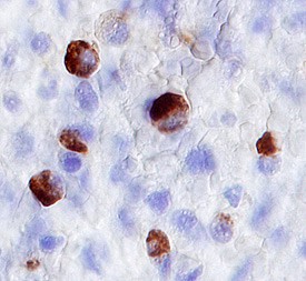 Myeloperoxidase/MPO in Human Spleen. Myeloperoxidase/MPO was detected in immersion fixed paraffin-embedded sections of human spleen using 3 µg/mL Goat Anti-Human/Mouse Myeloperoxidase/MPO Antigen Affinity-purified Polyclonal Antibody (Catalog # AF3667) overnight at 4 °C. Before incubation with the primary antibody tissue was subjected to heat-induced epitope retrieval using Antigen Retrieval Reagent-Basic (Catalog # CTS013). Tissue was stained with the Anti-Goat HRP-DAB Cell & Tissue Staining Kit (brown; Catalog # CTS008) and counterstained with hematoxylin (blue). View our protocol for Chromogenic IHC Staining of Paraffin-embedded Tissue Sections.
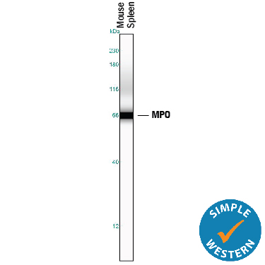 Detection of Mouse Myeloperoxidase/MPO by Simple WesternTM. Simple Western lane view shows lysates of mouse spleen tissue, loaded at 0.2 mg/mL. A specific band was detected for Myeloperoxidase/MPO at approximately 67 kDa (as indicated) using 5 µg/mL of Goat Anti-Human/Mouse Myeloperoxidase/MPO Antigen Affinity-purified Polyclonal Antibody (Catalog # AF3667) followed by 1:50 dilution of HRP-conjugated Anti-Goat IgG Secondary Antibody (Catalog # HAF109). This experiment was conducted under reducing conditions and using the12-230 kDa separation system.
Product References
Transcriptomic analysis of host immune and cell death responses associated with - Transcriptomic analysis of host immune and cell death responses associated with
Le Goffic R, Leymarie O, Chevalier C, Rebours E, Da Costa B, Vidic J, Descamps D, Sallenave JM, Rauch M, Samson M, Delmas B. PLoS Pathog. 2011 Aug;7(8):e1002202.
Tissue inhibitor of metalloproteinases 3 regulates resolution of inflammation - Tissue inhibitor of metalloproteinases 3 regulates resolution of inflammation
Gill SE, Huizar I, Bench EM, Sussman SW, Wang Y, Khokha R, Parks WC. Am J Pathol. 2010 Jan;176(1):64-73.