Human Phospho-EGF R/ErbB1 (Y1173) Affinity Purified PAb, Rabbit IgG
| Name | Human Phospho-EGF R/ErbB1 (Y1173) Affinity Purified PAb, Rabbit IgG |
|---|---|
| Supplier | R&D Systems |
| Catalog | AF1095-SP |
| Host | Rabbit |
| Clonality | Polyclonal |
| Isotype | IgG |
| Applications | Simple Western WB FC IHC ICC/IF |
| Species Reactivities | Human |
| Antigen | Phosphopeptide containing human EGF R Y1173 site |
| Description | Rabbit Polyclonal |
| Gene | EGFR |
| Conjugate | Unconjugated |
| Supplier Page | Shop |
Product images
 Detection of Human Phospho-EGF R/ErbB1 (Y1173) by Western Blot. Western blot shows lysates of A431 human epithelial carcinoma cell line untreated(‑) or treated (+) with 100 µM pervanadate (PV) for 10 minutes. PVDF membrane was probed with 0.2 µg/mL of Rabbit Anti-Human Phospho-EGF R/ErbB1 (Y1173) Antigen Affinity-purified Polyclonal Antibody, followed by HRP-conjugated Anti-Rabbit IgG Secondary Antibody (Catalog # HAF008). A specific band was detected for Phospho-EGF R/ErbB1 (Y1173) at approximately 185 kDa (as indicated). This experiment was conducted under reducing conditions and using Immunoblot Buffer Group 1.
 Detection of Phospho-EGF R/ErbB1 (Y1173) in A431 Human Cell Line by Flow Cytometry. A431 human epithelial carcinoma cells were untreated (open histogram), or treated for 5 minutes with 100 ng/mL Recombinant Human EGF (Catalog # 236-EG; filled histogram) then stained with Rabbit Anti-Human Phospho-EGF R/ErbB1 (Y1173) Antigen Affinity‑purified Polyclonal Antibody (Catalog # AF1095), followed by Phycoerythrin-conjugated Anti-Rabbit IgG Secondary Antibody (Catalog # F0110). To facilitate intracellular staining, cells were fixed with paraformaldehyde and permeabilized with saponin.
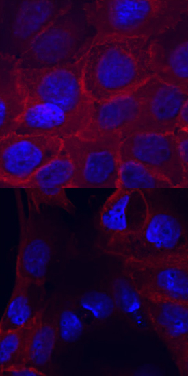 Phospho-EGF R/ErbB1 (Y1173) in A431 Human Cell Line. EGF R/ErbB1 phosphorylated at Y1173 was detected in immersion fixed A431 human epithelial carcinoma cell line untreated (lower panel) or treated (upper panel) with pervanadate using Rabbit Anti-Human Phospho-EGF R/ErbB1 (Y1173) Antigen Affinity-purified Polyclonal Antibody (Catalog # AF1095) at 10 µg/mL for 3 hours at room temperature. Cells were stained using the NorthernLights™ 557-conjugated Anti-Rabbit IgG Secondary Antibody (red; Catalog # NL004) and counterstained with DAPI (blue). View our protocol for Fluorescent ICC Staining of Cells on Coverslips.
 Phospho-EGF R/ErbB1 (Y1173) in Mouse Embryo. EGF R/ErbB1 phosphorylated at Y1173 was detected in immersion fixed frozen sections of mouse embryo using Rabbit Anti-Human Phospho-EGF R/ErbB1 (Y1173) Antigen Affinity-purified Polyclonal Antibody (Catalog # AF1095) at 15 µg/mL overnight at 4 °C. Tissue was stained using the Anti-Rabbit HRP-DAB Cell & Tissue Staining Kit (brown; Catalog # CTS005) and counterstained with hematoxylin (blue). Lower panel shows a lack of labeling if primary antibodies are omitted and tissue is stained only with secondary antibody followed by incubation with detection reagents. View our protocol for Chromogenic IHC Staining of Frozen Tissue Sections.
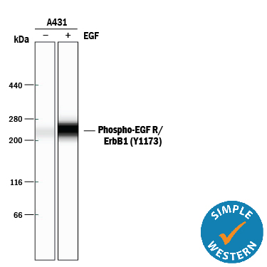 Detection of Human Phospho-EGF R/ErbB1 (Y1173) by Simple WesternTM. Simple Western lane view shows lysates of A431 human epithelial carcinoma cell line untreated (-) or treated (+) with 10 ng/mL Recombinant Human EGF (Catalog # 236-EG) for 5 minutes, loaded at 0.2 mg/mL. A specific band was detected for Phospho-EGF R/ErbB1 (Y1173) at approximately 265 kDa (as indicated) using 2 µg/mL of Rabbit Anti-Human Phospho-EGF R/ErbB1 (Y1173) Antigen Affinity-purified Polyclonal Antibody (Catalog # AF1095). This experiment was conducted under reducing conditions and using the 66-440 kDa separation system.
Product References
Protein C is an autocrine growth factor for human skin keratinocytes. - Protein C is an autocrine growth factor for human skin keratinocytes.
Xue M, Campbell D, Jackson CJ. J Biol Chem. 2007 May 4;282(18):13610-6. Epub 2007 Feb 9.
Repulsion of cerebellar granule neurons by chondroitin sulfate proteoglycans is - Repulsion of cerebellar granule neurons by chondroitin sulfate proteoglycans is
Kaneko M, Kubo T, Hata K, Yamaguchi A, Yamashita T. Neurosci Lett. 2007 Aug 9;423(1):62-7. Epub 2007 Jul 5.