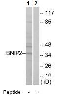
All lanes : Anti-BNIP2 antibody (ab74820) at 1/500 dilutionLane 1 : extracts from Jurkat cellsLane 2 : extracts from Jurkat cells with immunizing peptide at 10 µgLysates/proteins at 30 µg per lane.
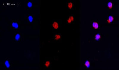
Central Panel: ab74820 staining BNIP2 in human PMN cells by ICC/IF (Immunocytochemistry/immunofluorescence). Cells were fixed with paraformaldehyde, permeabilized with 0.1% TritonX-100 + 2% BSA in PBS for 15 minutes and blocked with 2% BSA for 1 hour. Samples were incubated with primary antibody 1/100 in blocking buffer for 4 hours at 37°C. An Alexa Fluor® 568-conjugated Goat polyclonal to rabbit IgG, dilution 1/250, was used as secondary antibody. Left-hand panel: Nuclei counterstained with DAPI (blue).Right-hand panel: OverlaySee Abreview
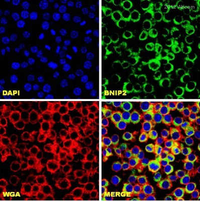
ab74820 staining BNIP2 (green) in Mouse RAW 264.7 cells by ICC (Immunocytochemistry). Cells were fixed with paraformaldehyde, permeabilized in 0.1% Triton X-100 in 2% BSA for 15 minutes and blocked with 2% BSA for 1 hour at 22°C. Samples were incubated with primary antibody (1/250 in PBS + 2% BSA) for 16 hours at 4°C. An Alexa Fluor®488-conjugated Chicken anti-rabbit IgG polyclonal (H&L) (1/750) was used as the secondary antibody. WGA (red) = Wheat Germ AgglutininSee Abreview