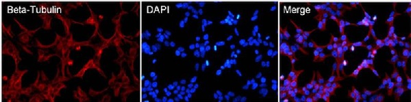
Immunofluorescent analysis of Beta-Tubulin (red) in HEK293T cells. Cells fixed in 4% formaldehyde were permeabilized and blocked with 1X PBS containing 5% BSA and 0.3% Triton X-100 for 1 hour at room temperature. Cells were probed with a Beta-Tubulin monoclonal antibody (ab173839) at a dilution of 1:100 overnight at 4ºC in 1X PBS containing 1% BSA and 0.3% Triton X-100, washed with 1X PBS, and incubated with a fluorophore-conjugated goat anti-mouse IgG secondary antibody at a dilution of 1:200 for 1 hour at room temperature. Nuclei (blue) were stained with DAPI. Images were taken at 40X magnification.
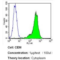
Flow cytometry analysis of Beta Tubulin in CEM cells (green) compared to an isotype control (blue). Cells were harvested, adjusted to a concentration of 1-5x10^6 cells/ml, fixed with 2% paraformaldehyde and washed with PBS. Cells were blocked with a 2% solution of BSA-PBS for 30 min at room temperature and incubated with a Beta Tubulin loading control antibody (ab173839) at a dilution of 1 ug/test for 40 min at room temperature. Cells were then incubated for 40 min at room temperature in the dark using a Dylight 488-conjugated secondary antibody and re-suspended in PBS for FACS analysis.
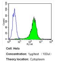
Flow cytometry analysis of Beta Tubulin in Hela cells (green) compared to an isotype control (blue). Cells were harvested, adjusted to a concentration of 1-5x10^6 cells/ml, fixed with 2% paraformaldehyde and washed with PBS. Cells were blocked with a 2% solution of BSA-PBS for 30 min at room temperature and incubated with a Beta Tubulin loading control antibody (ab173839) at a dilution of 1 ug/test for 40 min at room temperature. Cells were then incubated for 40 min at room temperature in the dark using a Dylight 488-conjugated secondary antibody and re-suspended in PBS for FACS analysis.
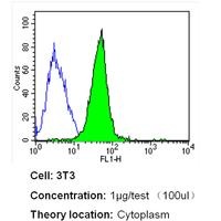
Flow cytometry analysis of Beta Tubulin in NIH-3T3 cells (green) compared to an isotype control (blue). Cells were harvested, adjusted to a concentration of 1-5x10^6 cells/ml, fixed with 2% paraformaldehyde and washed with PBS. Cells were blocked with a 2% solution of BSA-PBS for 30 min at room temperature and incubated with a Beta Tubulin loading control antibody (ab173839) at a dilution of 1 ug/test for 40 min at room temperature. Cells were then incubated for 40 min at room temperature in the dark using a Dylight 488-conjugated secondary antibody and re-suspended in PBS for FACS analysis.
![All lanes : Anti-beta Tubulin antibody [BT7R] (Biotin) (ab173839) at 1/1000 dilutionLane 1 : HeLa cell lysateLane 2 : 293T cell lysateLane 3 : A431 cell lysateLane 4 : COS7 cell lysateLane 5 : C2C12 cell lysateLane 6 : NRK cell lysateLysates/proteins at 50 µg per lane.SecondaryStreptavidin-HRP at 1/20000 dilutiondeveloped using the ECL technique](http://www.bioprodhub.com/system/product_images/ab_products/2/sub_1/14272_ab173839-197658-ab173839.jpg)
All lanes : Anti-beta Tubulin antibody [BT7R] (Biotin) (ab173839) at 1/1000 dilutionLane 1 : HeLa cell lysateLane 2 : 293T cell lysateLane 3 : A431 cell lysateLane 4 : COS7 cell lysateLane 5 : C2C12 cell lysateLane 6 : NRK cell lysateLysates/proteins at 50 µg per lane.SecondaryStreptavidin-HRP at 1/20000 dilutiondeveloped using the ECL technique