Anti-beta Actin antibody - Loading Control
| Name | Anti-beta Actin antibody - Loading Control |
|---|---|
| Supplier | Abcam |
| Catalog | ab13822 |
| Prices | $400.00 |
| Sizes | 100 µg |
| Host | Chicken |
| Clonality | Polyclonal |
| Isotype | IgY |
| Applications | IHC-P ICC/IF ICC/IF WB |
| Species Reactivities | Mouse, Rat, Human, Hamster, Rabbit, Chicken, Bovine, Dog, Xenopus, Fish |
| Antigen | Synthetic peptide derived from residues 1 - 100 of Human beta Actin |
| Description | Chicken Polyclonal |
| Gene | ACTB |
| Conjugate | Unconjugated |
| Supplier Page | Shop |
Product images
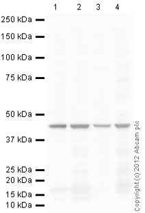 All lanes : Anti-beta Actin antibody - Loading Control (ab13822) at 1 µg/mlLane 1 : HeLa (Human epithelial carcinoma cell line) Whole Cell Lysate Lane 2 : NIH 3T3 (Mouse embryonic fibroblast cell line) Whole Cell Lysate Lane 3 : Liver (Rat) Tissue LysateLane 4 : CHO-K1 cell lysate Whole Cell Lysate Lysates/proteins at 10 µg per lane.SecondaryGoat polyclonal Secondary Antibody to Chicken IgY - H&L (HRP) at 1/3000 dilutiondeveloped using the ECL techniquePerformed under reducing conditions.
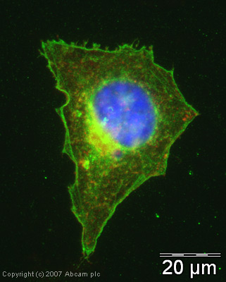 ICC/IF image of ab13822 stained human HeLa cells. The cells were methanol fixed (5 min), permabilised in TBS-T (20 min) and incubated with the antibody (ab13822, 5µg/ml) for 1h at room temperature. 1%BSA / 10% normal goat serum / 0.3M glycine was used to quench autofluorescence and block non-specific protein-protein interactions. The secondary antibody (green) was Alexa Fluor® 488 goat anti-chicken IgG (H+L) used at a 1/1000 dilution for 1h. Alexa Fluor® 594 WGA was used to label plasma membranes (red). DAPI was used to stain the cell nuclei (blue).
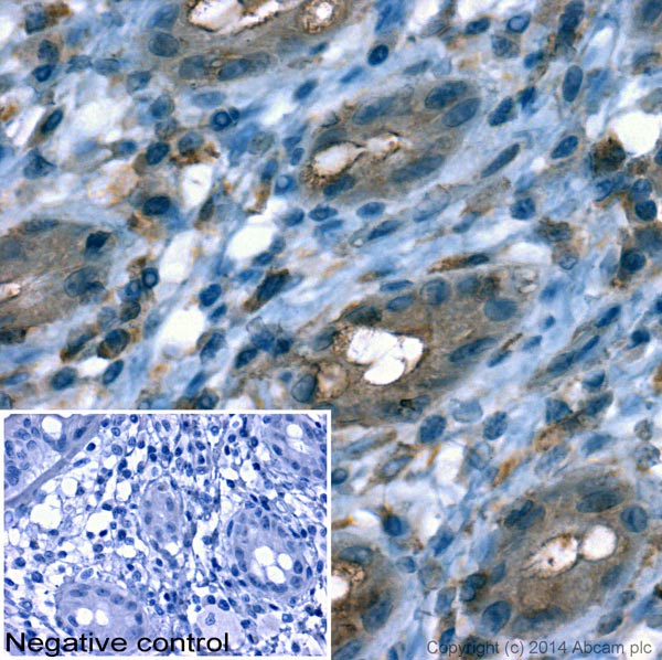 IHC image of beta Actin staining in THuman normal colon formalin fixed paraffin embedded tissue section*. The section was pre-treated using pressure cooker heat mediated antigen retrieval with sodium citrate buffer (pH6) for 30mins. The section was incubated with ab13822, 10µg/ml overnight at +4°C. A goat-anti chicken HRP secondary antibody (Ab6877, 1/500 dilution) was used for 1hr at room temperature. The section was counterstained with haematoxylin and mounted with DPX.*Tissue obtained from the Human Research Tissue Bank, supported by the NIHR Cambridge Biomedical Research Centre
Product References
Expression, functional, and structural analysis of proteins critical for otoconia - Expression, functional, and structural analysis of proteins critical for otoconia
Xu Y, Zhang H, Yang H, Zhao X, Lovas S, Lundberg YW. Dev Dyn. 2010 Oct;239(10):2659-73.
Regional hippocampal differences in AKT survival signaling across the lifespan: - Regional hippocampal differences in AKT survival signaling across the lifespan:
Jackson TC, Rani A, Kumar A, Foster TC. Cell Death Differ. 2009 Mar;16(3):439-48.
Multi-step pericellular proteolysis controls the transition from individual to - Multi-step pericellular proteolysis controls the transition from individual to
Wolf K, Wu YI, Liu Y, Geiger J, Tam E, Overall C, Stack MS, Friedl P. Nat Cell Biol. 2007 Aug;9(8):893-904. Epub 2007 Jul 8.