Anti-beta Actin antibody [mAbcam 8226]
| Name | Anti-beta Actin antibody [mAbcam 8226] |
|---|---|
| Supplier | Abcam |
| Catalog | ab8226 |
| Prices | $404.00 |
| Sizes | 100 µg |
| Host | Mouse |
| Clonality | Monoclonal |
| Isotype | IgG1 |
| Clone | mAbcam 8226 |
| Applications | ICC/IF ICC/IF ICC/IF FC IHC-F IHC-P IHC-F IP WB |
| Species Reactivities | Mouse, Rat, Rabbit, Horse, Chicken, Bovine, Dog, Human, Pig, Zebrafish, Monkey, Hamster, Sheep, Guinea Pig |
| Antigen | Synthetic peptide conjugated to KLH derived from within residues 1 - 100 of Human beta Actin |
| Description | Mouse Monoclonal |
| Gene | ACTB |
| Conjugate | Unconjugated |
| Supplier Page | Shop |
Product images
![Lanes 1 - 3 : Anti-beta Actin antibody [mAbcam 8226] (ab8226) at 1 µg/ml (5% BSA BLOCK)Lanes 4 - 6 : Anti-beta Actin antibody [mAbcam 8226] (ab8226) at 1 µg/ml (5% MILK BLOCK)Lane 1 : HeLa (Human epithelial carcinoma cell line) Whole Cell Lysate Lane 2 : Jurkat (Human T cell lymphoblast-like cell line) Whole Cell Lysate Lane 3 : NIH 3T3 (Mouse embryonic fibroblast cell line) Whole Cell LysateLane 4 : HeLa (Human epithelial carcinoma cell line) Whole Cell Lysate Lane 5 : Jurkat (Human T cell lymphoblast-like cell line) Whole Cell Lysate Lane 6 : NIH 3T3 (Mouse embryonic fibroblast cell line) Whole Cell LysateLysates/proteins at 10 µg per lane.SecondaryGoat polyclonal to Mouse IgG - H&L - Pre-Adsorbed (HRP) at 1/3000 dilutionPerformed under reducing conditions.](http://www.bioprodhub.com/system/product_images/ab_products/2/sub_1/13680_beta-Actin-Primary-antibodies-ab8226-27.jpg) Lanes 1 - 3 : Anti-beta Actin antibody [mAbcam 8226] (ab8226) at 1 µg/ml (5% BSA BLOCK)Lanes 4 - 6 : Anti-beta Actin antibody [mAbcam 8226] (ab8226) at 1 µg/ml (5% MILK BLOCK)Lane 1 : HeLa (Human epithelial carcinoma cell line) Whole Cell Lysate Lane 2 : Jurkat (Human T cell lymphoblast-like cell line) Whole Cell Lysate Lane 3 : NIH 3T3 (Mouse embryonic fibroblast cell line) Whole Cell LysateLane 4 : HeLa (Human epithelial carcinoma cell line) Whole Cell Lysate Lane 5 : Jurkat (Human T cell lymphoblast-like cell line) Whole Cell Lysate Lane 6 : NIH 3T3 (Mouse embryonic fibroblast cell line) Whole Cell LysateLysates/proteins at 10 µg per lane.SecondaryGoat polyclonal to Mouse IgG - H&L - Pre-Adsorbed (HRP) at 1/3000 dilutionPerformed under reducing conditions.
Lanes 1 - 3 : Anti-beta Actin antibody [mAbcam 8226] (ab8226) at 1 µg/ml (5% BSA BLOCK)Lanes 4 - 6 : Anti-beta Actin antibody [mAbcam 8226] (ab8226) at 1 µg/ml (5% MILK BLOCK)Lane 1 : HeLa (Human epithelial carcinoma cell line) Whole Cell Lysate Lane 2 : Jurkat (Human T cell lymphoblast-like cell line) Whole Cell Lysate Lane 3 : NIH 3T3 (Mouse embryonic fibroblast cell line) Whole Cell LysateLane 4 : HeLa (Human epithelial carcinoma cell line) Whole Cell Lysate Lane 5 : Jurkat (Human T cell lymphoblast-like cell line) Whole Cell Lysate Lane 6 : NIH 3T3 (Mouse embryonic fibroblast cell line) Whole Cell LysateLysates/proteins at 10 µg per lane.SecondaryGoat polyclonal to Mouse IgG - H&L - Pre-Adsorbed (HRP) at 1/3000 dilutionPerformed under reducing conditions.
![All lanes : Anti-beta Actin antibody [mAbcam 8226] (ab8226) at 1/1000 dilutionLane 1 : A431 (Human epithelial carcinoma cell line) Whole Cell LysateLane 2 : HEK293 (Human embryonic kidney cell line) Whole Cell LysateLane 3 : NIH 3T3 (Mouse embryonic fibroblast cell line) Whole Cell LysateLane 4 : PC12 (Rat adrenal pheochromocytoma cell line) Whole Cell LysateLysates/proteins at 20 µg per lane.SecondaryGoat Anti-Mouse IgG H&L (Alexa Fluor® 790) (ab175783) at 1/10000 dilution](http://www.bioprodhub.com/system/product_images/ab_products/2/sub_1/13681_ab8226-219013-FWBab8226DS.jpg) All lanes : Anti-beta Actin antibody [mAbcam 8226] (ab8226) at 1/1000 dilutionLane 1 : A431 (Human epithelial carcinoma cell line) Whole Cell LysateLane 2 : HEK293 (Human embryonic kidney cell line) Whole Cell LysateLane 3 : NIH 3T3 (Mouse embryonic fibroblast cell line) Whole Cell LysateLane 4 : PC12 (Rat adrenal pheochromocytoma cell line) Whole Cell LysateLysates/proteins at 20 µg per lane.SecondaryGoat Anti-Mouse IgG H&L (Alexa Fluor® 790) (ab175783) at 1/10000 dilution
All lanes : Anti-beta Actin antibody [mAbcam 8226] (ab8226) at 1/1000 dilutionLane 1 : A431 (Human epithelial carcinoma cell line) Whole Cell LysateLane 2 : HEK293 (Human embryonic kidney cell line) Whole Cell LysateLane 3 : NIH 3T3 (Mouse embryonic fibroblast cell line) Whole Cell LysateLane 4 : PC12 (Rat adrenal pheochromocytoma cell line) Whole Cell LysateLysates/proteins at 20 µg per lane.SecondaryGoat Anti-Mouse IgG H&L (Alexa Fluor® 790) (ab175783) at 1/10000 dilution
 Immunofluorescence using ab8226 at 5ug/ml incubated for 1 hour on Rat Colon Cancer cells. Cells were fixed with ice-cold methanol for 5 mins, then for all following steps, permeabilised in TBS-T for 30 mins, blocked with 5% BSA for 30 mins and then washed in TBS-T. Secondary antibody was Alexa Fluor 488 goat anti-mouse IgG at 1/1000 incubated for 1 hour. Cells were counterstained with DAPI. Image at 400X magnification. All incubations were at room temperature.The beta actin fibres can be seen arrayed around the edge of the cells.
![All lanes : Anti-beta Actin antibody [mAbcam 8226] (ab8226) at 1 µg/mlLane 1 : HeLa (Human epithelial carcinoma cell line) Whole Cell LysateLane 2 : NIH 3T3 (Mouse embryonic fibroblast cell line) Whole Cell LysateLane 3 : PC12 (Rat adrenal pheochromocytoma cell line) Whole Cell LysateLysates/proteins at 10 µg per lane.SecondaryGoat Anti-Mouse IgG H&L (HRP) preadsorbed (ab97040) at 1/50000 dilutiondeveloped using the ECL techniquePerformed under reducing conditions.](http://www.bioprodhub.com/system/product_images/ab_products/2/sub_1/13683_ab8226-219469-WBAP17122472.jpg) All lanes : Anti-beta Actin antibody [mAbcam 8226] (ab8226) at 1 µg/mlLane 1 : HeLa (Human epithelial carcinoma cell line) Whole Cell LysateLane 2 : NIH 3T3 (Mouse embryonic fibroblast cell line) Whole Cell LysateLane 3 : PC12 (Rat adrenal pheochromocytoma cell line) Whole Cell LysateLysates/proteins at 10 µg per lane.SecondaryGoat Anti-Mouse IgG H&L (HRP) preadsorbed (ab97040) at 1/50000 dilutiondeveloped using the ECL techniquePerformed under reducing conditions.
All lanes : Anti-beta Actin antibody [mAbcam 8226] (ab8226) at 1 µg/mlLane 1 : HeLa (Human epithelial carcinoma cell line) Whole Cell LysateLane 2 : NIH 3T3 (Mouse embryonic fibroblast cell line) Whole Cell LysateLane 3 : PC12 (Rat adrenal pheochromocytoma cell line) Whole Cell LysateLysates/proteins at 10 µg per lane.SecondaryGoat Anti-Mouse IgG H&L (HRP) preadsorbed (ab97040) at 1/50000 dilutiondeveloped using the ECL techniquePerformed under reducing conditions.
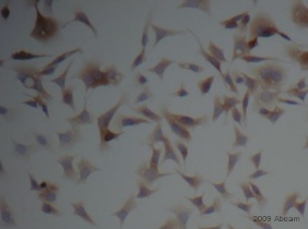 ab8226 staining beta Actin in human AGS cells by Immunocytochemistry. Cells were fixed with formaldehyde, permeabilized with 0.025% Triton-X in TBS and blocking with 5% serum was performed for 1 hour at 230C. Samples were incubated with primary antibody (1/500) for 1 hour at 23°C. An HRP®-conjugated goat polyclonal to mouse IgG was used undiluted as secondary antibody.See Abreview
![Anti-beta Actin antibody [mAbcam 8226] (ab8226) at 0.5 µg/ml + HeLa cell lysateSecondaryGoat polyclonal to mouse IgG H&L (HRP) at 1/5000 dilutionPerformed under non-reducing conditions.](http://www.bioprodhub.com/system/product_images/ab_products/2/sub_1/13685_ab8226_3.jpg) Anti-beta Actin antibody [mAbcam 8226] (ab8226) at 0.5 µg/ml + HeLa cell lysateSecondaryGoat polyclonal to mouse IgG H&L (HRP) at 1/5000 dilutionPerformed under non-reducing conditions.
Anti-beta Actin antibody [mAbcam 8226] (ab8226) at 0.5 µg/ml + HeLa cell lysateSecondaryGoat polyclonal to mouse IgG H&L (HRP) at 1/5000 dilutionPerformed under non-reducing conditions.
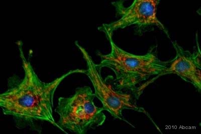 ab8226 staining beta Actin in human gastric epithelial AGS cells by Immunocytochemistry/ Immunofluorescence. Cells were fixed in formaldehyde. Permeabilization and blocking was carried out using 5% BSA containing 0.025% Triton X in TBS for 1 hour at 23°C. Primary antibody was used at 5µg/ml for 1 hour at 23°C. An Alexa Fluor 488® conjugated goat polyclonal to mouse IgG was used as the secondary antibody at a 1/1000 dilution.See Abreview
![All lanes : Anti-beta Actin antibody [mAbcam 8226] (ab8226) at 1 µg/mlLane 1 : HeLa (Human epithelial carcinoma cell line) Whole Cell LysateLane 2 : Jurkat (Human T cell lymphoblast-like cell line) Whole Cell LysateLane 3 : A431 (Human epithelial carcinoma cell line) Whole Cell LysateLane 4 : HEK293 (Human embryonic kidney cell line) Whole Cell LysateLane 5 : HepG2 (Human hepatocellular liver carcinoma cell line) Whole Cell LysateLysates/proteins at 20 µg per lane.SecondaryGoat Anti-Rabbit IgG H&L (HRP) (ab97051) at 1/10000 dilutiondeveloped using the ECL techniquePerformed under reducing conditions.](http://www.bioprodhub.com/system/product_images/ab_products/2/sub_1/13687_beta-Actin-Primary-antibodies-ab8226-97.jpg) All lanes : Anti-beta Actin antibody [mAbcam 8226] (ab8226) at 1 µg/mlLane 1 : HeLa (Human epithelial carcinoma cell line) Whole Cell LysateLane 2 : Jurkat (Human T cell lymphoblast-like cell line) Whole Cell LysateLane 3 : A431 (Human epithelial carcinoma cell line) Whole Cell LysateLane 4 : HEK293 (Human embryonic kidney cell line) Whole Cell LysateLane 5 : HepG2 (Human hepatocellular liver carcinoma cell line) Whole Cell LysateLysates/proteins at 20 µg per lane.SecondaryGoat Anti-Rabbit IgG H&L (HRP) (ab97051) at 1/10000 dilutiondeveloped using the ECL techniquePerformed under reducing conditions.
All lanes : Anti-beta Actin antibody [mAbcam 8226] (ab8226) at 1 µg/mlLane 1 : HeLa (Human epithelial carcinoma cell line) Whole Cell LysateLane 2 : Jurkat (Human T cell lymphoblast-like cell line) Whole Cell LysateLane 3 : A431 (Human epithelial carcinoma cell line) Whole Cell LysateLane 4 : HEK293 (Human embryonic kidney cell line) Whole Cell LysateLane 5 : HepG2 (Human hepatocellular liver carcinoma cell line) Whole Cell LysateLysates/proteins at 20 µg per lane.SecondaryGoat Anti-Rabbit IgG H&L (HRP) (ab97051) at 1/10000 dilutiondeveloped using the ECL techniquePerformed under reducing conditions.
![Lane 1 : Anti-beta Actin antibody [mAbcam 8226] (ab8226) at 1/1000 dilutionLane 2 : Anti-beta Actin antibody [mAbcam 8226] (ab8226) at 1/10000 dilutionLanes 3 - 4 : Anti-beta Actin antibody [mAbcam 8226] (ab8226) at 1/500 dilutionLane 1 : HeLa Cell lysateLane 2 : HeLa Cell lysateLane 3 : 293 Cell lysateLane 4 : 3T3 Mouse Cell lysateLysates/proteins at 20 µg per lane.SecondaryRabbit Anti-Mouse IgG H&L (HRP) (ab6728) at 1/5000 dilutionPerformed under reducing conditions.](http://www.bioprodhub.com/system/product_images/ab_products/2/sub_1/13688_ab8226_2.jpg) Lane 1 : Anti-beta Actin antibody [mAbcam 8226] (ab8226) at 1/1000 dilutionLane 2 : Anti-beta Actin antibody [mAbcam 8226] (ab8226) at 1/10000 dilutionLanes 3 - 4 : Anti-beta Actin antibody [mAbcam 8226] (ab8226) at 1/500 dilutionLane 1 : HeLa Cell lysateLane 2 : HeLa Cell lysateLane 3 : 293 Cell lysateLane 4 : 3T3 Mouse Cell lysateLysates/proteins at 20 µg per lane.SecondaryRabbit Anti-Mouse IgG H&L (HRP) (ab6728) at 1/5000 dilutionPerformed under reducing conditions.
Lane 1 : Anti-beta Actin antibody [mAbcam 8226] (ab8226) at 1/1000 dilutionLane 2 : Anti-beta Actin antibody [mAbcam 8226] (ab8226) at 1/10000 dilutionLanes 3 - 4 : Anti-beta Actin antibody [mAbcam 8226] (ab8226) at 1/500 dilutionLane 1 : HeLa Cell lysateLane 2 : HeLa Cell lysateLane 3 : 293 Cell lysateLane 4 : 3T3 Mouse Cell lysateLysates/proteins at 20 µg per lane.SecondaryRabbit Anti-Mouse IgG H&L (HRP) (ab6728) at 1/5000 dilutionPerformed under reducing conditions.
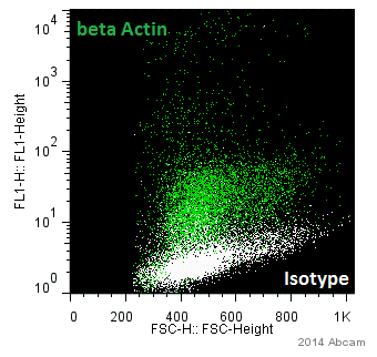 ab8226 staining beta Actin in human LGE neural progenitors by Flow Cytometry. Cells were paraformaldehyde and permeabilized. The sample was incubated with the primary antibody (1/200) for 1 hour at 20°C. An Alexa Fluor® 488-conjugated goat anti-mouse IgG1 (1/800) was used as the secondary antibody.Gating Strategy: Isotype population (shown in white).See Abreview
Product References
Lipopolysaccharide activates Toll-like receptor 4 (TLR4)-mediated NF-kappaB - Lipopolysaccharide activates Toll-like receptor 4 (TLR4)-mediated NF-kappaB
Guijarro-Munoz I, Compte M, Alvarez-Cienfuegos A, Alvarez-Vallina L, Sanz L. J Biol Chem. 2014 Jan 24;289(4):2457-68.
Promyelocytic leukemia protein is a cell-intrinsic factor inhibiting parvovirus - Promyelocytic leukemia protein is a cell-intrinsic factor inhibiting parvovirus
Mitchell AM, Hirsch ML, Li C, Samulski RJ. J Virol. 2014 Jan;88(2):925-36.
Calcineurin suppresses AMPK-dependent cytoprotective autophagy in cardiomyocytes - Calcineurin suppresses AMPK-dependent cytoprotective autophagy in cardiomyocytes
He H, Liu X, Lv L, Liang H, Leng B, Zhao D, Zhang Y, Du Z, Chen X, Li S, Lu Y, Shan H. Cell Death Dis. 2014 Jan 16;5:e997.
Epidermal growth factor upregulates Skp2/Cks1 and p27(kip1) in human extrahepatic - Epidermal growth factor upregulates Skp2/Cks1 and p27(kip1) in human extrahepatic
Kim JY, Kim HJ, Park JH, Park DI, Cho YK, Sohn CI, Jeon WK, Kim BI, Kim DH, Chae SW, Sohn JH. World J Gastroenterol. 2014 Jan 21;20(3):755-73.
Medaka villin 1-like protein (VILL) is associated with the formation of - Medaka villin 1-like protein (VILL) is associated with the formation of
Kang CK, Lee TH. Front Zool. 2014 Jan 13;11(1):2.
Increased microRNA-34c abundance in Alzheimer's disease circulating blood plasma. - Increased microRNA-34c abundance in Alzheimer's disease circulating blood plasma.
Bhatnagar S, Chertkow H, Schipper HM, Yuan Z, Shetty V, Jenkins S, Jones T, Wang E. Front Mol Neurosci. 2014 Feb 4;7:2.
A fluorescence-coupled assay for gamma aminobutyric acid (GABA) reveals metabolic - A fluorescence-coupled assay for gamma aminobutyric acid (GABA) reveals metabolic
Ippolito JE, Piwnica-Worms D. PLoS One. 2014 Feb 13;9(2):e88667.
A defect in the CLIP1 gene (CLIP-170) can cause autosomal recessive intellectual - A defect in the CLIP1 gene (CLIP-170) can cause autosomal recessive intellectual
Larti F, Kahrizi K, Musante L, Hu H, Papari E, Fattahi Z, Bazazzadegan N, Liu Z, Banan M, Garshasbi M, Wienker TF, Ropers HH, Galjart N, Najmabadi H. Eur J Hum Genet. 2015 Mar;23(3):331-6.
Chronic exposure to type-I IFN under lymphopenic conditions alters CD4 T cell - Chronic exposure to type-I IFN under lymphopenic conditions alters CD4 T cell
Le Saout C, Hasley RB, Imamichi H, Tcheung L, Hu Z, Luckey MA, Park JH, Durum SK, Smith M, Rupert AW, Sneller MC, Lane HC, Catalfamo M. PLoS Pathog. 2014 Mar 6;10(3):e1003976.
Calreticulin from suboolemmal vesicles affects membrane regulation of polyspermy. - Calreticulin from suboolemmal vesicles affects membrane regulation of polyspermy.
Saavedra MD, Mondejar I, Coy P, Betancourt M, Gonzalez-Marquez H, Jimenez-Movilla M, Aviles M, Romar R. Reproduction. 2014 Feb 5;147(3):369-78.