
Ab36585, at a 1/100 - 1/500 dilution, staining Bcl-10 in paraffin embedded human liver carcinoma tissue by immunohistochemistry with DAB staining.

Ab36585, at a 1/100 - 1/500 dilution, staining Bcl-10 in paraffin embedded human breast carcinoma tissue by immunohistochemistry with DAB staining.
![All lanes : Anti-Bcl10 antibody [4F8E8H8] (ab36585) at 1/500 dilutionLane 1 : Cell lysates prepared from NIH/3T3 cellsLane 2 : Cell lysates prepared from Hela cellsLane 3 : Cell lysates prepared from MCF-7 cellsLane 4 : Cell lysates prepared from Jurkat cellsLysates/proteins at 100 µg per lane.SecondaryHRP-conjugated Goat polyclonal to mouse IgG1](http://www.bioprodhub.com/system/product_images/ab_products/2/sub_1/13095_Bcl10-Primary-antibodies-ab36585-1.jpg)
All lanes : Anti-Bcl10 antibody [4F8E8H8] (ab36585) at 1/500 dilutionLane 1 : Cell lysates prepared from NIH/3T3 cellsLane 2 : Cell lysates prepared from Hela cellsLane 3 : Cell lysates prepared from MCF-7 cellsLane 4 : Cell lysates prepared from Jurkat cellsLysates/proteins at 100 µg per lane.SecondaryHRP-conjugated Goat polyclonal to mouse IgG1
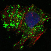
ab36585 at 1/1000 dilution staining Bcl10 in human Hela cells by Immunocytochemistry/ Immunofluorescence. Bcl10 show green staining in image and actin filament stained red with Alexa Fluor®555 conjugated secondary. Nuclei stained blue with DRAQ5 fluorescent DNA dye.
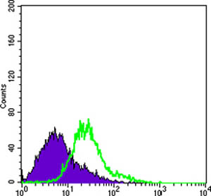
Flow cytometric analysis of Hela cells using ab36585 at a 1/200 dilution (green) and negative control (purple).