![All lanes : Anti-Aurora B antibody [mAbcam 3609] (ab3609) at 1 µg/mlLane 1 : Hek293 Whole Cell LysateLane 2 : Jurkat Whole Cell LysateLane 3 : HeLa Whole Cell LysateLane 4 : HeLa Whole Cell Lysate- Nocodozole StimulatedLysates/proteins at 20 µg per lane.SecondaryGoat polyclonal to Mouse IgG - H&L - Pre-Absorbed (HRP) at 1/3000 dilutiondeveloped using the ECL techniquePerformed under reducing conditions.](http://www.bioprodhub.com/system/product_images/ab_products/2/sub_1/11694_ab3609-240249-ID943Ab3609Ap18317043mindatasheetimage.jpg)
All lanes : Anti-Aurora B antibody [mAbcam 3609] (ab3609) at 1 µg/mlLane 1 : Hek293 Whole Cell LysateLane 2 : Jurkat Whole Cell LysateLane 3 : HeLa Whole Cell LysateLane 4 : HeLa Whole Cell Lysate- Nocodozole StimulatedLysates/proteins at 20 µg per lane.SecondaryGoat polyclonal to Mouse IgG - H&L - Pre-Absorbed (HRP) at 1/3000 dilutiondeveloped using the ECL techniquePerformed under reducing conditions.
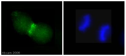
SKN-SH cells were fixed in 4% paraformaldehyde for 10 mins, permeabilized in PBS-0.5% Triton X-100 for 5 mins and incubated for 30 minutes with ab3609 (1/50 dilution). The slides were blocked with 4mg/ml BSA. The slides were rinsed once in PBS-Triton (0.1%), twice in PBS then incubated with the secondary antibody for 30 mins. The DNA is stained with DAPI (blue). Staining with ab3609 can be seen in green.

Image courtesy of Human Protein AtlasParaffin embedded human liver tumor tissue was incubated with ab3609 (1/35 dilution) for 30 mins at room temperature. Antigen retrieval was performed by heat induction in citrate buffer pH 6.ab3609 was tested in a tissue microarray (TMA) containing a wide range of normal and cancer tissues as well as a cell microarray consisting of a range of commonly used, well characterised human cell lines. Further images can be found at www.proteinatlas.org
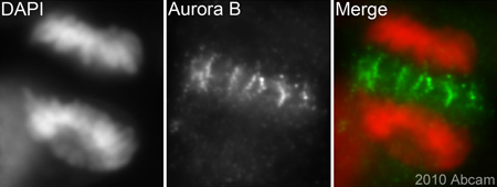
ab3609 (1/200) staining Aurora B in HeLa cells (green). Cells were fixed in paraformaldehyde, permeabilised with 0.5% Triton X-100/PBS and counterstained with DAPI in order to highlight the nucleus/ chromosomes (red). For further experimental details please refer to Abreview.See Abreview