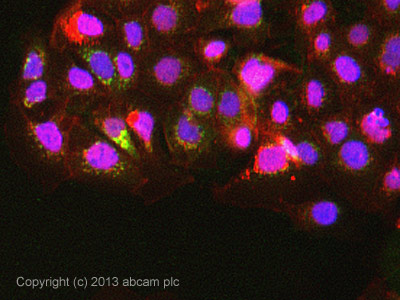
ICC/IF image of ab155982 stained CaCo2 cells. The cells were 4% formaldehyde fixed (10 min) and then incubated in 1%BSA / 10% normal goat serum / 0.3M glycine in 0.1% PBS-Tween for 1h to permeabilise the cells and block non-specific protein-protein interactions. The cells were then incubated with the antibody ab155982 at 10µg/ml overnight at +4°C. The secondary antibody (green) was DyLight® 488 goat anti- rabbit (ab96899) IgG (H+L) used at a 1/250 dilution for 1h. Alexa Fluor® 594 WGA was used to label plasma membranes (red) at a 1/200 dilution for 1h. DAPI was used to stain the cell nuclei (blue) at a concentration of 1.43µM.
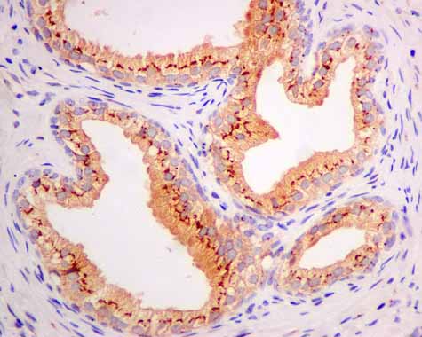
ab155982 showing +ve staining in Human normal prostate.
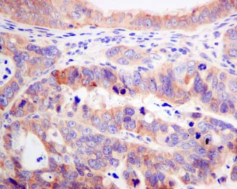
ab155982 showing +ve staining in Human gastric adenocarcinoma.
ab155982 showing +ve staining in Human endometrial carcinoma.
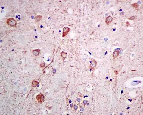
ab155982 showing +ve staining in Human normal brain.
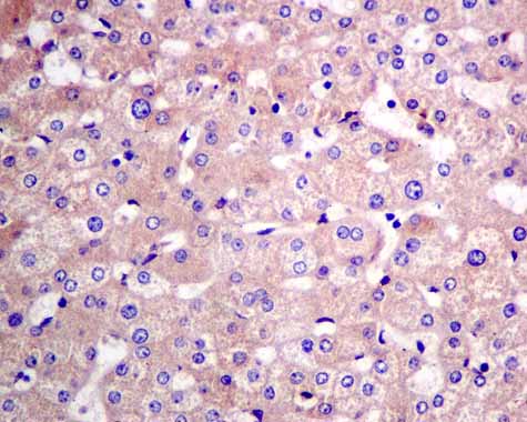
ab155982 showing +ve staining in Human normal liver.
![All lanes : Anti-ARL 1 antibody [EPR10595] (ab155982) at 1/1000 dilutionLane 1 : HepG2 cell lysateLane 2 : MCF7 cell lysateLane 3 : HeLa cell lysateLane 4 : K562 cell lysateLysates/proteins at 10 µg per lane.SecondaryHRP labeled goat anti-rabbit IgG at 1/2000 dilution](http://www.bioprodhub.com/system/product_images/ab_products/2/sub_1/9454_ARL-1-Primary-antibodies-ab155982-2.jpg)
All lanes : Anti-ARL 1 antibody [EPR10595] (ab155982) at 1/1000 dilutionLane 1 : HepG2 cell lysateLane 2 : MCF7 cell lysateLane 3 : HeLa cell lysateLane 4 : K562 cell lysateLysates/proteins at 10 µg per lane.SecondaryHRP labeled goat anti-rabbit IgG at 1/2000 dilution
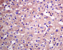
Immunohistochemical analysis of paraffin-embedded Human liver tissue labeling ARL 1 with ab155982 at 1/50 dilution.
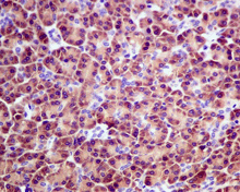
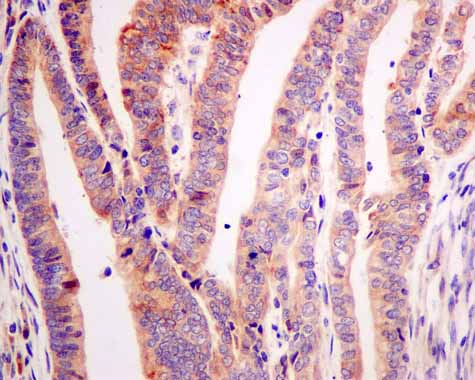
Immunohistochemical analysis of formalin-fixed, paraffin-embedded Human pancreas tissue labeling ARL 1 with ab155982 at 1/50 dilution