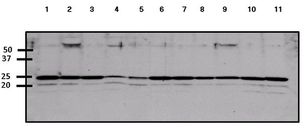
All lanes : Anti-ARF6 antibody (ab155929) at 1/1000 dilutionLane 1 : HeLa whole cell lysateLane 2 : HepG2 whole cell lysateLane 3 : 293T whole cell lysateLane 4 : A431 whole cell lysateLane 5 : Jurkat whole cell lysateLane 6 : MCF7 whole cell lysateLane 7 : U2OS whole cell lysateLane 8 : K562 whole cell lysateLane 9 : COS7 whole cell lysateLane 10 : MEF whole cell lysateLane 11 : 3T3L1 whole cell lysateLysates/proteins at 50 µg per lane.SecondaryGoat anti-rabbit HRP conjugated antibody at 1/10000 dilutiondeveloped using the ECL technique
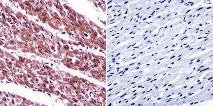
Immunohistochemical analysis of deparaffinized Human heart tissue labelling ARF6 with ab155929 at 1/20. The panel on the right shows the tissue without primary antibody incubation (negative control).
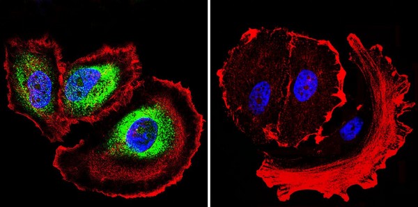
Immunofluorescent analysis of formaldehyde fixed MCF7 cells labeling ARF6 with ab155929 at 1/20 (green). The panel on the right shows cells without primary antibody incubation (negative control). Both panels show F-Actin staining with Phalloidin (red) and nuclei with DAPI (blue).
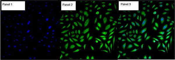
Immunofluorescent analysis of formalin fixed, permeabilized HeLa cells labeling ARF6 with ab155929 at 1/100 (green). Panel 1 shows cell nuclei staining with Hoechst 33342 only (blue), Panel 2 with ab155929 only and Panel 3 with Hoeschst 33342 merged with ab155929.