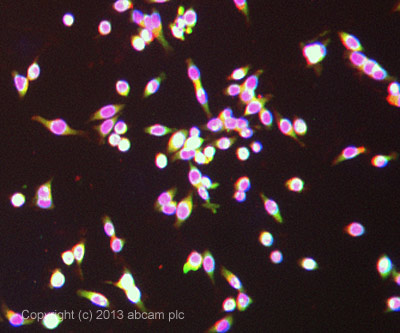
ab42673 stained LoVo cells. The cells were 4% formaldehyde fixed (10 min) and then incubated in 1%BSA / 10% normal goat serum / 0.3M glycine in 0.1% PBS-Tween for 1h to permeabilise the cells and block non-specific protein-protein interactions. The cells were then incubated with the antibody ab42673 at 5µg/ml overnight at +4°C. The secondary antibody (green) was DyLight® 488 goat anti- rabbit (ab96899) IgG (H+L) used at a 1/250 dilution for 1h. Alexa Fluor® 594 WGA was used to label plasma membranes (red) at a 1/200 dilution for 1h. DAPI was used to stain the cell nuclei (blue) at a concentration of 1.43µM.
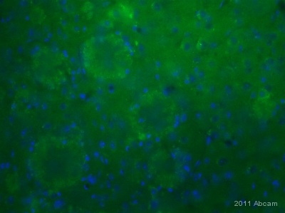
IHC-FoFr image of Apolipoprotein J stained APP tg mouse cortex using ab42673. The animal were perfuse with 4% PFA.The sections were incubated in 10% normal donkey serum in 0.1% PBS- and triton X100 for 1h to permeabilise the cells and block non-specific protein-protein interactions. The sections were then incubated with the antibody (ab42673, 1µg/ml) overnight at +4°C. The secondary antibody (green) was Alexa Fluor® 488 donkey anti-rabbit IgG (H+L) used at a 1/1000 dilution for 1h. DAPI was used to stain the cell nuclei (blue) at a concentration of 1.43µM. See Abreview

Anti-Apolipoprotein J antibody (ab42673) at 1 µg/ml + Testis (Human) Tissue Lysate - adult normal tissue (ab30257) at 10 µgSecondaryIRDye 680 Conjugated Goat Anti-Rabbit IgG (H+L) at 1/10000 dilutionPerformed under reducing conditions.