Anti-APAF1 antibody [E38]
| Name | Anti-APAF1 antibody [E38] |
|---|---|
| Supplier | Abcam |
| Catalog | ab32372 |
| Prices | $401.00 |
| Sizes | 100 µl |
| Host | Rabbit |
| Clonality | Monoclonal |
| Isotype | IgG |
| Clone | E38 |
| Applications | ICC/IF ICC/IF WB IHC-P |
| Species Reactivities | Mouse, Human |
| Antigen | Synthetic peptide (the amino acid sequence is considered to be commercially sensitive) corresponding to Human APAF1 (N terminal) |
| Description | Rabbit Monoclonal |
| Gene | APAF1 |
| Conjugate | Unconjugated |
| Supplier Page | Shop |
Product images
![Anti-APAF1 antibody [E38] (ab32372) at 1/1000 dilution (purified) + Mouse spleen tissue lysate at 20 µgSecondaryHRP goat anti-rabbit (H+L) at 1/1000 dilution](http://www.bioprodhub.com/system/product_images/ab_products/2/sub_1/7924_ab32372-5-ab32372WB2.jpg) Anti-APAF1 antibody [E38] (ab32372) at 1/1000 dilution (purified) + Mouse spleen tissue lysate at 20 µgSecondaryHRP goat anti-rabbit (H+L) at 1/1000 dilution
Anti-APAF1 antibody [E38] (ab32372) at 1/1000 dilution (purified) + Mouse spleen tissue lysate at 20 µgSecondaryHRP goat anti-rabbit (H+L) at 1/1000 dilution
![All lanes : Anti-APAF1 antibody [E38] (ab32372) at 1/1000 dilution (purified)Lane 1 : HeLa cell lysateLane 2 : MCF7 cell lysateLane 3 : HT-1080 cell lysateLysates/proteins at 20 µg per lane.SecondaryHRP goat anti-rabbit (H+L) at 1/1000 dilution](http://www.bioprodhub.com/system/product_images/ab_products/2/sub_1/7925_ab32372-4-ab32372WB1.jpg) All lanes : Anti-APAF1 antibody [E38] (ab32372) at 1/1000 dilution (purified)Lane 1 : HeLa cell lysateLane 2 : MCF7 cell lysateLane 3 : HT-1080 cell lysateLysates/proteins at 20 µg per lane.SecondaryHRP goat anti-rabbit (H+L) at 1/1000 dilution
All lanes : Anti-APAF1 antibody [E38] (ab32372) at 1/1000 dilution (purified)Lane 1 : HeLa cell lysateLane 2 : MCF7 cell lysateLane 3 : HT-1080 cell lysateLysates/proteins at 20 µg per lane.SecondaryHRP goat anti-rabbit (H+L) at 1/1000 dilution
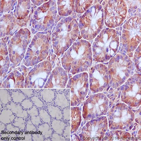 Immunohistochemical staining of paraffin embedded mouse colon with purified ab32372 at a working dilution of 1/250. The secondary antibody used is ab97051, a HRP-conjugated goat anti-rabbit IgG (H+L), at a dilution of 1/500. The sample is counter-stained with hematoxylin. Antigen retrieval was perfomed using Tris-EDTA buffer, pH 9.0. PBS was used instead of the primary antibody as the negative control, and is shown in the inset.
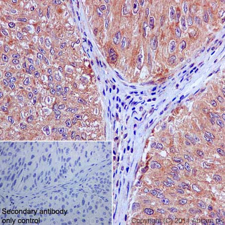 Immunohistochemical staining of paraffin embedded human bladder cancer with purified ab32372 at a working dilution of 1/250. The secondary antibody used is ab97051, a HRP-conjugated goat anti-rabbit IgG (H+L), at a dilution of 1/500. The sample is counter-stained with hematoxylin. Antigen retrieval was perfomed using Tris-EDTA buffer, pH 9.0. PBS was used instead of the primary antibody as the negative control, and is shown in the inset.
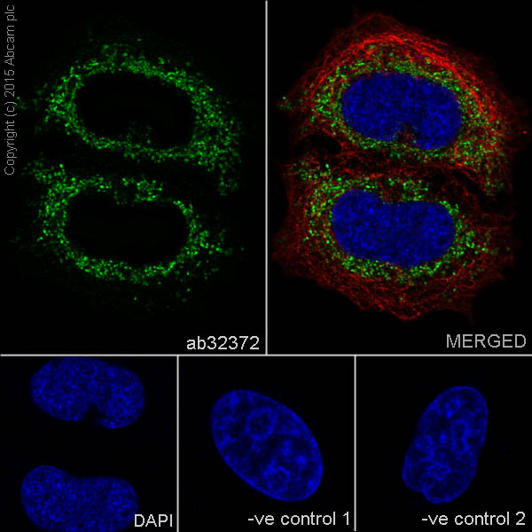 Immunofluorescence staining of HeLa cells with purified ab32372 at a working dilution of 1 in 100, counter-stained with DAPI. Tubulin was stained with mouse anti-tubulin at a dilution of 1/1000 (ab7291) and Alexa Fluor® 594 goat anti-mouse at a dilution of 1/500 (ab150120) . The secondary antibody was ab150077 Alexa Fluor® 488 goat anti rabbit, used at a dilution of 1 in 500. The cells were fixed in 4% PFA and permeabilized using 0.1% Triton X 100. The negative controls are shown in the bottom middle and right hand panels - for the first negative control, purified ab323722 was used at a dilution of 1/200 followed by an Alexa Fluor® 555 goat anti-mouse antibody at a dilution of 1/500 and for the second negative control mouse primary antibody (ab7291) and anti-rabbit secondary antibody (ab15007) were used.
![Anti-APAF1 antibody [E38] (ab32372) at 1/500 dilution (unpurified) + HeLa cell lysate](http://www.bioprodhub.com/system/product_images/ab_products/2/sub_1/7929_ab32372_1.jpg) Anti-APAF1 antibody [E38] (ab32372) at 1/500 dilution (unpurified) + HeLa cell lysate
Anti-APAF1 antibody [E38] (ab32372) at 1/500 dilution (unpurified) + HeLa cell lysate
 Unpurified ab32372, at a dilution of 1/50, staining APAF1 in paraffin embedded stomach adenocarcinoma by Immunohistochemistry.Ab32372, at a dilution of 1/50, staining APAF1 in paraffin embedded stomach adenocarcinoma by Immunohistochemistry.
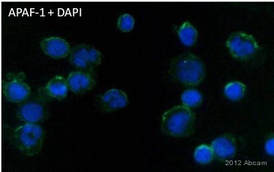 Unpurified ab32372 staining APAF1 in human leukocytes by Immunocytochemistry/ Immunofluorescence.Cells were fixed in formaldehyde, permeabilized using 0.3% Triton X-100, blocked with 2% serum for 2 hours at 25°C, and then incubated with ab32372 at a 1/250 dilution for 9 hours at 4°C. The secondary used was an Alexa-Fluor 488 conjugated goat anti-rabbit polyclonal used at a 1/500 dilution.See Abreview
Product References
ELL inhibits E2F1 transcriptional activity by enhancing E2F1 deacetylation via - ELL inhibits E2F1 transcriptional activity by enhancing E2F1 deacetylation via
Zhang W, Ji W, Liu X, Ouyang G, Xiao W. Mol Cell Biol. 2014 Feb;34(4):765-75.
MicroRNA-23a/b and microRNA-27a/b suppress Apaf-1 protein and alleviate - MicroRNA-23a/b and microRNA-27a/b suppress Apaf-1 protein and alleviate
Chen Q, Xu J, Li L, Li H, Mao S, Zhang F, Zen K, Zhang CY, Zhang Q. Cell Death Dis. 2014 Mar 20;5:e1132.
p63 is an alternative p53 repressor in melanoma that confers chemoresistance and - p63 is an alternative p53 repressor in melanoma that confers chemoresistance and
Matin RN, Chikh A, Chong SL, Mesher D, Graf M, Sanza' P, Senatore V, Scatolini M, Moretti F, Leigh IM, Proby CM, Costanzo A, Chiorino G, Cerio R, Harwood CA, Bergamaschi D. J Exp Med. 2013 Mar 11;210(3):581-603.
MiR-155 inhibits the sensitivity of lung cancer cells to cisplatin via negative - MiR-155 inhibits the sensitivity of lung cancer cells to cisplatin via negative
Zang YS, Zhong YF, Fang Z, Li B, An J. Cancer Gene Ther. 2012 Nov;19(11):773-8.
A short caspase-3 isoform inhibits chemotherapy-induced apoptosis by blocking - A short caspase-3 isoform inhibits chemotherapy-induced apoptosis by blocking
Vegran F, Boidot R, Solary E, Lizard-Nacol S. PLoS One. 2011;6(12):e29058.
The Bcr-Abl kinase inhibitor INNO-406 induces autophagy and different modes of - The Bcr-Abl kinase inhibitor INNO-406 induces autophagy and different modes of
Kamitsuji Y, Kuroda J, Kimura S, Toyokuni S, Watanabe K, Ashihara E, Tanaka H, Yui Y, Watanabe M, Matsubara H, Mizushima Y, Hiraumi Y, Kawata E, Yoshikawa T, Maekawa T, Nakahata T, Adachi S. Cell Death Differ. 2008 Nov;15(11):1712-22.