
ab6330 staining AMACR by immunohistochemistry on paraffin-embedded human normal prostate tissue (left, light staining) and on prostate adenocarcinoma using DAB staining (right)
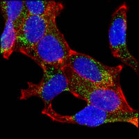
ab63340 at 1/1000 dilution staining AMACR in LnCap cells by Immunocytochemistry/ Immunofluorescence. An Alexa Fluor® 488 conjugated goat anti-mouse {IgG (H+C)} was used as secondary. Actin filaments were stained red with DY-554 phalloidin. Nuclei were stained blue with DRAQ5 fluorescent DNA dye.
![All lanes : Anti-AMACR antibody [2A10F3] (ab63340) at 1/2000 dilutionLane 1 : Cell lysates prepared from Jurkat cellsLane 2 : Cell lysates prepared from LnCaP cellsLysates/proteins at 100 µg per lane.SecondaryHRP-conjugated Goat polyclonal to mouse IgG1](http://www.bioprodhub.com/system/product_images/ab_products/2/sub_1/6309_AMACR-Primary-antibodies-ab63340-2.jpg)
All lanes : Anti-AMACR antibody [2A10F3] (ab63340) at 1/2000 dilutionLane 1 : Cell lysates prepared from Jurkat cellsLane 2 : Cell lysates prepared from LnCaP cellsLysates/proteins at 100 µg per lane.SecondaryHRP-conjugated Goat polyclonal to mouse IgG1
![Overlay histogram showing PC3 cells stained with ab63340 (red line). The cells were fixed with 80% methanol (5 min) and then permeabilized with 0.1% PBS-Tween for 20 min. The cells were then incubated in 1x PBS / 10% normal goat serum / 0.3M glycine to block non-specific protein-protein interactions. The cells were then incubated with the antibody (ab63340, 1µg/1x106 cells) for 30 min at 22ºC. The secondary antibody used was DyLight® 488 goat anti-mouse IgG (H+L) (ab96879) at 1/500 dilution for 30 min at 22ºC. Isotype control antibody (black line) was mouse IgG2b [PLPV219] (ab91366, 2µg/1x106 cells) used under the same conditions. Acquisition of >5,000 events was performed. This antibody gave a positive signal in PC3 cells fixed with 4% paraformaldehyde (10 min)/permeabilized with 0.1% PBS-Tween for 20 min used under the same conditions.](http://www.bioprodhub.com/system/product_images/ab_products/2/sub_1/6310_AMACR-Primary-antibodies-ab63340-3.jpg)
Overlay histogram showing PC3 cells stained with ab63340 (red line). The cells were fixed with 80% methanol (5 min) and then permeabilized with 0.1% PBS-Tween for 20 min. The cells were then incubated in 1x PBS / 10% normal goat serum / 0.3M glycine to block non-specific protein-protein interactions. The cells were then incubated with the antibody (ab63340, 1µg/1x106 cells) for 30 min at 22ºC. The secondary antibody used was DyLight® 488 goat anti-mouse IgG (H+L) (ab96879) at 1/500 dilution for 30 min at 22ºC. Isotype control antibody (black line) was mouse IgG2b [PLPV219] (ab91366, 2µg/1x106 cells) used under the same conditions. Acquisition of >5,000 events was performed. This antibody gave a positive signal in PC3 cells fixed with 4% paraformaldehyde (10 min)/permeabilized with 0.1% PBS-Tween for 20 min used under the same conditions.