Anti-alpha Tubulin antibody [EP1332Y] - Microtubule Marker
| Name | Anti-alpha Tubulin antibody [EP1332Y] - Microtubule Marker |
|---|---|
| Supplier | Abcam |
| Catalog | ab52866 |
| Prices | $404.00 |
| Sizes | 100 µl |
| Host | Rabbit |
| Clonality | Monoclonal |
| Isotype | IgG |
| Clone | EP1332Y |
| Applications | ICC/IF ICC/IF IHC WB IP FC IHC-P IHC-F |
| Species Reactivities | Mouse, Rat, Human, Pig, Drosophila |
| Antigen | Synthetic peptide (the amino acid sequence is considered to be commercially sensitive) (N terminal) |
| Description | Rabbit Monoclonal |
| Gene | TUBA4A |
| Conjugate | Unconjugated |
| Supplier Page | Shop |
Product images
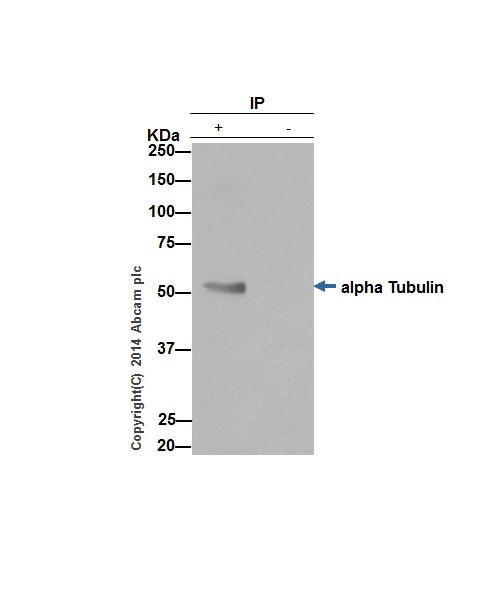 alpha Tubulin was immunoprecipitated from 1mg of HeLa whole cell extract using ab52866 at 1/30 dilution. Western blot was performed from the immunoprecipitate using ab52866 at 1/1000 dilution. Anti-Rabbit IgG (HRP), specific to the non-reduced form of IgG, was used as secondary antibody at 1/1500 dilution.Lane 1 Hela whole cell extract, Lane 2 PBS instead of whole cell extract.Blocking and dilution buffer and concentration: 5% NFDM/TBST.
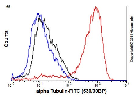 Flow cytometry analysis of 2% paraformaldehyde fixed HepG2 cells labeling alpha Tubulin with ab52866 at 1/130 dilution (red line). Secondary antibody used is a goat anti rabbit IgG (FITC) at 1/150 dilution. The isotype control is rabbit monoclonal IgG (black line). The unlabeled control is cells without incubation with primary and secondary antibodies (blue line).
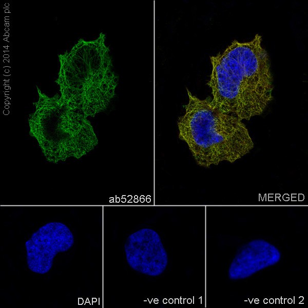 Immunofluorescent analysis of 4% paraformaldehyde-fixed HeLa cells labeling alpha Tubulin with ab52866 at 1/500 dilution. The cells were permeabilised with 0.1% Triton X-100. Anti-rabbit Alexa Fluor® 488 (ab150077) at 1/400 dilution was used as the secondary antibody (green). The confocal image shows microtubules staining on HeLa cell line. The nuclear counter stain is DAPI (blue). Tubulin is detected with ab7291 (anti-Tubulin mouse mAb) at 1/500 and anti-mouse AlexaFluor® 594 (ab150120) at 1/500 dilution (red).The negative controls are as follows:1. ab52866 at 1/500 dilution followed by anti-mouse AlexaFluor® 594 (ab150120) at 1/500 dilution.2. ab7291 (anti-Tubulin mouse mAb) at 1/500 dilution followed by anti-rabbit Alexa Fluor® 488 (ab150077) at 1/400 dilution.
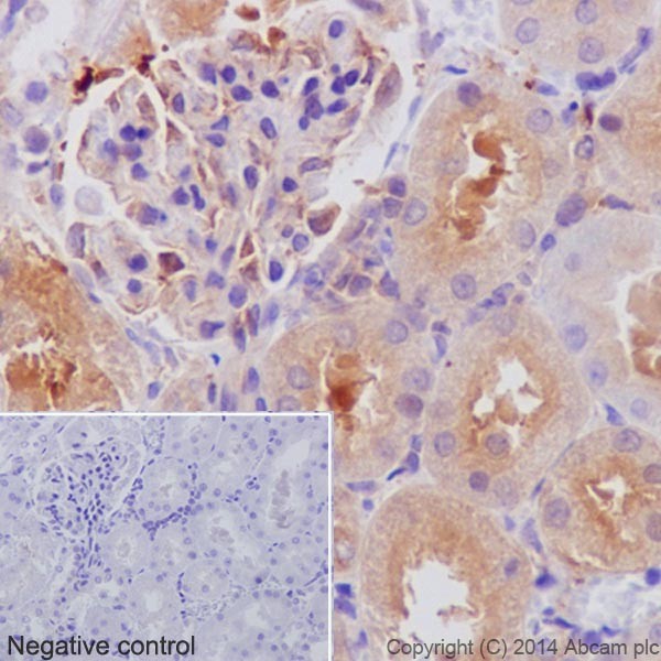
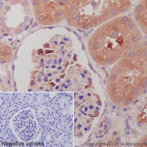 Immunohistochemistry analysis of paraffin-embedded Pig kidney tissue labeling alpha Tubulin with ab52866 at a 1/1000 dilution. Cytoplasmic staining on Pig kidney tubule and weak on glomerulus shown. Anti-Rabbit HRP (ab97051) used at a 1/100 dilution. Counter stained with Hematoxylin.Inset image: negative control obtained using PBS instead of ab52866, secondary antibody is Anti-Rabbit HRP (ab97051) at 1/100 dilution.
 Immunohistochemistry analysis of paraffin-embedded Rat kidney tissue labeling alpha Tubulin with ab52866 at a 1/1000 dilution. Cytoplasmic staining on Rat kidney tubule and weak on glomerulus shown. Secondary antibody Anti-Rabbit HRP (ab97051) used at a 1/500 dilution. Counter stained with Hematoxylin.Inset image: negative control obtained using PBS instead of ab52866, secondary antibody is Anti-Rabbit HRP (ab97051) at 1/500 dilution.
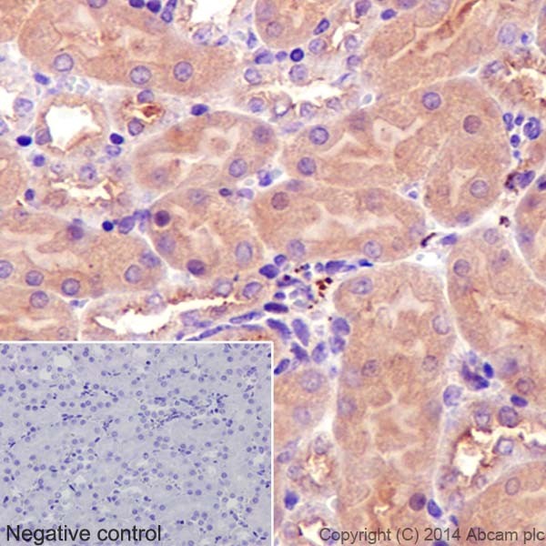 Immunohistochemistry analysis of paraffin-embedded Mouse kidney tissue labeling alpha Tubulin with ab52866 at a 1/1000 dilution. Cytoplasmic staining on Mouse kidney tubule shown. Secondary antibody Anti-Rabbit HRP (ab97051) used at a 1/500 dilution. Counter stained with Hematoxylin.Inset image: negative control obtained using PBS instead of ab52866, secondary antibody is Anti-Rabbit HRP (ab97051) at 1/500 dilution.
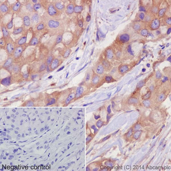 Immunohistochemistry analysis of paraffin-embedded Human breast cancer labeling alpha Tubulin with ab52866 at a 1/1000 dilution. Cytoplasmic staining on cancer cells shown. Secondary antibody ab97051 Goat Anti-Rabbit IgG H&L (HRP) used at a 1/500 dilution. Counter stained with Hematoxylin.Inset image: negative control obtained using PBS instead of ab52866, secondary antibody is Anti-Rabbit HRP (ab97051) at 1/500 dilution.
![Anti-alpha Tubulin antibody [EP1332Y] - Microtubule Marker (ab52866) at 1/50000 dilution + Rat brain lysates at 10 µgSecondaryAnti-Rabbit IgG (HRP), specific to the non-reduced form of IgG at 1/1000 dilution](http://www.bioprodhub.com/system/product_images/ab_products/2/sub_1/6102_ab52866-234662-ab52866WB5.jpg) Anti-alpha Tubulin antibody [EP1332Y] - Microtubule Marker (ab52866) at 1/50000 dilution + Rat brain lysates at 10 µgSecondaryAnti-Rabbit IgG (HRP), specific to the non-reduced form of IgG at 1/1000 dilution
Anti-alpha Tubulin antibody [EP1332Y] - Microtubule Marker (ab52866) at 1/50000 dilution + Rat brain lysates at 10 µgSecondaryAnti-Rabbit IgG (HRP), specific to the non-reduced form of IgG at 1/1000 dilution
![All lanes : Anti-alpha Tubulin antibody [EP1332Y] - Microtubule Marker (ab52866) at 1/5000 dilutionLane 1 : Mouse brain lysatesLane 2 : C6 whole cell lysatesLane 3 : Raw264.7 whole cell lysatesLane 4 : PC-12 whole cell lysatesLane 5 : NIH3T3 whole cell lysatesLysates/proteins at 10 µg per lane.SecondaryAnti-Rabbit IgG (HRP), specific to the non-reduced form of IgG at 1/1000 dilution](http://www.bioprodhub.com/system/product_images/ab_products/2/sub_1/6103_ab52866-234661-ab52866WB4.jpg) All lanes : Anti-alpha Tubulin antibody [EP1332Y] - Microtubule Marker (ab52866) at 1/5000 dilutionLane 1 : Mouse brain lysatesLane 2 : C6 whole cell lysatesLane 3 : Raw264.7 whole cell lysatesLane 4 : PC-12 whole cell lysatesLane 5 : NIH3T3 whole cell lysatesLysates/proteins at 10 µg per lane.SecondaryAnti-Rabbit IgG (HRP), specific to the non-reduced form of IgG at 1/1000 dilution
All lanes : Anti-alpha Tubulin antibody [EP1332Y] - Microtubule Marker (ab52866) at 1/5000 dilutionLane 1 : Mouse brain lysatesLane 2 : C6 whole cell lysatesLane 3 : Raw264.7 whole cell lysatesLane 4 : PC-12 whole cell lysatesLane 5 : NIH3T3 whole cell lysatesLysates/proteins at 10 µg per lane.SecondaryAnti-Rabbit IgG (HRP), specific to the non-reduced form of IgG at 1/1000 dilution
![All lanes : Anti-alpha Tubulin antibody [EP1332Y] - Microtubule Marker (ab52866) at 1/1000 dilutionLane 1 : HeLa (Human epithelial carcinoma cell line) Whole Cell LysateLane 2 : HEK293 (Human embryonic kidney cell line) Whole Cell LysateLane 3 : HepG2 (Human hepatocellular liver carcinoma cell line) Whole Cell LysateLane 4 : Caco 2 (Human colonic carcinoma cell line) Whole Cell LysateLane 5 : NIH 3T3 (Mouse embryonic fibroblast cell line) Whole Cell LysateLane 6 : PC12 (Rat adrenal pheochromocytoma cell line) Whole Cell LysateLysates/proteins at 20 µg per lane.SecondaryGoat Anti-Rabbit IgG H&L (Alexa Fluor® 790) (ab175781) at 1/10000 dilution](http://www.bioprodhub.com/system/product_images/ab_products/2/sub_1/6104_ab52866-221352-WBGR32556111.jpg) All lanes : Anti-alpha Tubulin antibody [EP1332Y] - Microtubule Marker (ab52866) at 1/1000 dilutionLane 1 : HeLa (Human epithelial carcinoma cell line) Whole Cell LysateLane 2 : HEK293 (Human embryonic kidney cell line) Whole Cell LysateLane 3 : HepG2 (Human hepatocellular liver carcinoma cell line) Whole Cell LysateLane 4 : Caco 2 (Human colonic carcinoma cell line) Whole Cell LysateLane 5 : NIH 3T3 (Mouse embryonic fibroblast cell line) Whole Cell LysateLane 6 : PC12 (Rat adrenal pheochromocytoma cell line) Whole Cell LysateLysates/proteins at 20 µg per lane.SecondaryGoat Anti-Rabbit IgG H&L (Alexa Fluor® 790) (ab175781) at 1/10000 dilution
All lanes : Anti-alpha Tubulin antibody [EP1332Y] - Microtubule Marker (ab52866) at 1/1000 dilutionLane 1 : HeLa (Human epithelial carcinoma cell line) Whole Cell LysateLane 2 : HEK293 (Human embryonic kidney cell line) Whole Cell LysateLane 3 : HepG2 (Human hepatocellular liver carcinoma cell line) Whole Cell LysateLane 4 : Caco 2 (Human colonic carcinoma cell line) Whole Cell LysateLane 5 : NIH 3T3 (Mouse embryonic fibroblast cell line) Whole Cell LysateLane 6 : PC12 (Rat adrenal pheochromocytoma cell line) Whole Cell LysateLysates/proteins at 20 µg per lane.SecondaryGoat Anti-Rabbit IgG H&L (Alexa Fluor® 790) (ab175781) at 1/10000 dilution
![Anti-alpha Tubulin antibody [EP1332Y] - Microtubule Marker (ab52866) at 1/50000 dilution + Human fetal kidney lysates at 10 µgSecondaryAnti-Rabbit IgG (HRP), specific to the non-reduced form of IgG at 1/1000 dilution](http://www.bioprodhub.com/system/product_images/ab_products/2/sub_1/6105_ab52866-234660-ab52866WB3.jpg) Anti-alpha Tubulin antibody [EP1332Y] - Microtubule Marker (ab52866) at 1/50000 dilution + Human fetal kidney lysates at 10 µgSecondaryAnti-Rabbit IgG (HRP), specific to the non-reduced form of IgG at 1/1000 dilution
Anti-alpha Tubulin antibody [EP1332Y] - Microtubule Marker (ab52866) at 1/50000 dilution + Human fetal kidney lysates at 10 µgSecondaryAnti-Rabbit IgG (HRP), specific to the non-reduced form of IgG at 1/1000 dilution
![Anti-alpha Tubulin antibody [EP1332Y] - Microtubule Marker (ab52866) at 1/5000 dilution + Pig skeletal muscle lysates at 20 µgSecondaryAnti-Rabbit IgG (HRP), specific to the non-reduced form of IgG at 1/1000 dilution](http://www.bioprodhub.com/system/product_images/ab_products/2/sub_1/6106_ab52866-234659-ab52866WB2.jpg) Anti-alpha Tubulin antibody [EP1332Y] - Microtubule Marker (ab52866) at 1/5000 dilution + Pig skeletal muscle lysates at 20 µgSecondaryAnti-Rabbit IgG (HRP), specific to the non-reduced form of IgG at 1/1000 dilution
Anti-alpha Tubulin antibody [EP1332Y] - Microtubule Marker (ab52866) at 1/5000 dilution + Pig skeletal muscle lysates at 20 µgSecondaryAnti-Rabbit IgG (HRP), specific to the non-reduced form of IgG at 1/1000 dilution
![All lanes : Anti-alpha Tubulin antibody [EP1332Y] - Microtubule Marker (ab52866) at 1/20000 dilutionLane 1 : HeLa whole cell lysateLane 2 : HepG2 whole cell lysateLane 3 : Jurkat whole cell lysateLane 4 : 293T whole cell lysateLysates/proteins at 20 µg per lane.SecondaryGoat Anti-Rabbit IgG, (H+L),Peroxidase conjugated at 1/1000 dilution](http://www.bioprodhub.com/system/product_images/ab_products/2/sub_1/6107_ab52866-234658-ab52866WB1.jpg) All lanes : Anti-alpha Tubulin antibody [EP1332Y] - Microtubule Marker (ab52866) at 1/20000 dilutionLane 1 : HeLa whole cell lysateLane 2 : HepG2 whole cell lysateLane 3 : Jurkat whole cell lysateLane 4 : 293T whole cell lysateLysates/proteins at 20 µg per lane.SecondaryGoat Anti-Rabbit IgG, (H+L),Peroxidase conjugated at 1/1000 dilution
All lanes : Anti-alpha Tubulin antibody [EP1332Y] - Microtubule Marker (ab52866) at 1/20000 dilutionLane 1 : HeLa whole cell lysateLane 2 : HepG2 whole cell lysateLane 3 : Jurkat whole cell lysateLane 4 : 293T whole cell lysateLysates/proteins at 20 µg per lane.SecondaryGoat Anti-Rabbit IgG, (H+L),Peroxidase conjugated at 1/1000 dilution
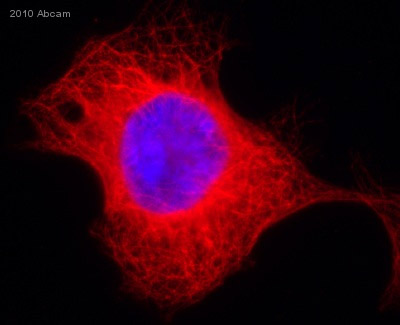 ab52866 staining alpha Tubulin in 293 Human embryonic kidney cells by ICC/IF (Immunocytochemistry/immunofluorescence). Cells were fixed with paraformaldehyde and blocked with 10% serum for 2 hours at 23°C. Samples were incubated with primary antibody (1/200 in 0.5% saponin) for 2 hours at 23°C. An Alexa Fluor®555-conjugated Goat anti-rabbit IgG polyclonal (1/500) was used as the secondary antibody. Nuclei were counterstained with DAPI.See Abreview
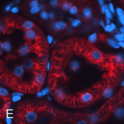 Immunohistochemical analysis of rat kidney tubule tissue, staining alpha Tubulin with ab52866.
Product References
GPR30 activation decreases anxiety in the open field test but not in the elevated - GPR30 activation decreases anxiety in the open field test but not in the elevated
Anchan D, Clark S, Pollard K, Vasudevan N. Brain Behav. 2014 Jan;4(1):51-9.
The H3K4 methyltransferase Setd1a is first required at the epiblast stage, - The H3K4 methyltransferase Setd1a is first required at the epiblast stage,
Bledau AS, Schmidt K, Neumann K, Hill U, Ciotta G, Gupta A, Torres DC, Fu J, Kranz A, Stewart AF, Anastassiadis K. Development. 2014 Mar;141(5):1022-35.
The NFL-TBS.40-63 anti-glioblastoma peptide disrupts microtubule and - The NFL-TBS.40-63 anti-glioblastoma peptide disrupts microtubule and
Rivalin R, Lepinoux-Chambaud C, Eyer J, Savagner F. PLoS One. 2014 Jun 4;9(6):e98473.
Drosophila Brahma complex remodels nucleosome organizations in multiple aspects. - Drosophila Brahma complex remodels nucleosome organizations in multiple aspects.
Shi J, Zheng M, Ye Y, Li M, Chen X, Hu X, Sun J, Zhang X, Jiang C. Nucleic Acids Res. 2014 Sep;42(15):9730-9.
Microtubule-associated type II protein kinase A is important for neurite - Microtubule-associated type II protein kinase A is important for neurite
Huang YA, Kao JW, Tseng DT, Chen WS, Chiang MH, Hwang E. PLoS One. 2013 Aug 13;8(8):e73890.
Sumoylation at chromatin governs coordinated repression of a transcriptional - Sumoylation at chromatin governs coordinated repression of a transcriptional
Neyret-Kahn H, Benhamed M, Ye T, Le Gras S, Cossec JC, Lapaquette P, Bischof O, Ouspenskaia M, Dasso M, Seeler J, Davidson I, Dejean A. Genome Res. 2013 Oct;23(10):1563-79.
Activity of LaSOM 65, a monastrol-derived compound, against glioblastoma - Activity of LaSOM 65, a monastrol-derived compound, against glioblastoma
Stuepp CS, Figueiro F, Mendes FB, Braganhol E, Bernardi A, Frozza RL, Salbego CG, Canto RF, Russowsky D, Eifler-Lima VL, Battastini AM. Anticancer Res. 2013 Oct;33(10):4463-8.
Activation of the G-protein coupled receptor 30 (GPR30) has different effects on - Activation of the G-protein coupled receptor 30 (GPR30) has different effects on
Hart D, Nilges M, Pollard K, Lynn T, Patsos O, Shiel C, Clark SM, Vasudevan N. Steroids. 2014 Mar;81:49-56.
Heparanase-induced GEF-H1 signaling regulates the cytoskeletal dynamics of brain - Heparanase-induced GEF-H1 signaling regulates the cytoskeletal dynamics of brain
Ridgway LD, Wetzel MD, Ngo JA, Erdreich-Epstein A, Marchetti D. Mol Cancer Res. 2012 Jun;10(6):689-702.
Morphological changes in diabetic kidney are associated with increased - Morphological changes in diabetic kidney are associated with increased
Akimoto Y, Miura Y, Toda T, Wolfert MA, Wells L, Boons GJ, Hart GW, Endo T, Kawakami H. Clin Proteomics. 2011 Sep 21;8(1):15.