Anti-alpha Tubulin antibody
| Name | Anti-alpha Tubulin antibody |
|---|---|
| Supplier | Abcam |
| Catalog | ab18251 |
| Prices | $400.00 |
| Sizes | 50 µg |
| Host | Rabbit |
| Clonality | Polyclonal |
| Isotype | IgG |
| Applications | ICC/IF ICC/IF IHC-P WB |
| Species Reactivities | Mouse, Rat, Chicken, Bovine, Human, Drosophila, Deer, Monkey, Hamster |
| Antigen | Synthetic peptide conjugated to KLH derived from within residues 400 to the C-terminus of Human alpha Tubulin |
| Description | Rabbit Polyclonal |
| Gene | TUBA4A |
| Conjugate | Unconjugated |
| Supplier Page | Shop |
Product images
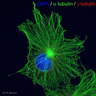
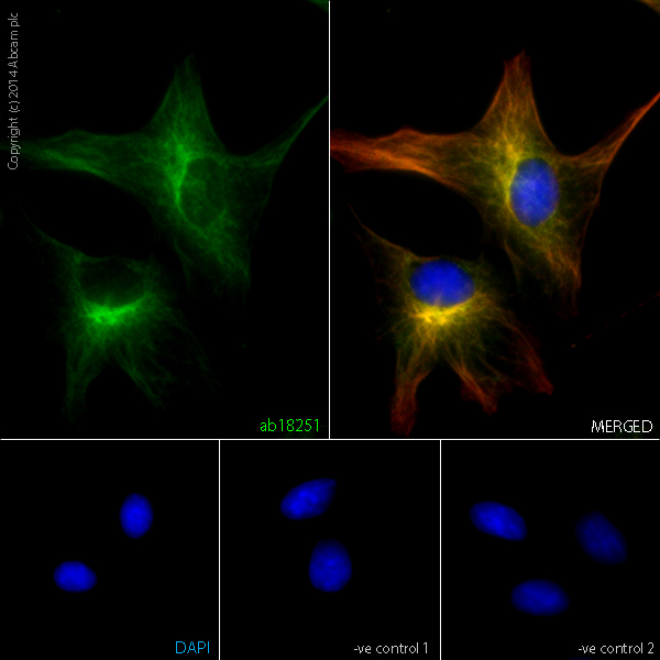 ab18251 staining alpha-Tubulin in SV40 cells. The cells were fixed with 100% methanol (5min) and then blocked in 1% BSA/10% normal goat serum/0.3M glycine in 0.1%PBS-Tween for 1h. The cells were then incubated with ab18251 at 1μl/ml and ab7291 at 1µg/ml overnight at +4°C, followed by a further incubation at room temperature for 1h with an anti-rabbit AlexaFluor® 488 (ab150081) at 2 μg/ml (shown in green) and anti-mouse AlexaFluor® 594 (ab150120) at 2 μg/ml (shown in pseudo color red). Nuclear DNA was labelled in blue with DAPI.Negative controls: 1– Rabbit primary antibody and anti-mouse secondary antibody; 2 – Mouse primary antibody and anti-rabbit secondary antibody. Controls 1 and 2 indicate that there is no unspecific reaction between primary and secondary antibodies used.
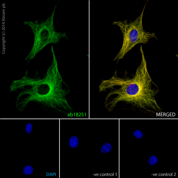 ab18251 staining alpha-Tubulin in NIH3T3 cells. The cells were fixed with 100% methanol (5min) and then blocked in 1% BSA/10% normal goat serum/0.3M glycine in 0.1%PBS-Tween for 1h. The cells were then incubated with ab18251 at 1μl/ml and ab7291 at 1µg/ml overnight at +4°C, followed by a further incubation at room temperature for 1h with an anti-rabbit AlexaFluor® 488 (ab150081) at 2 μg/ml (shown in green) and anti-mouse AlexaFluor® 594 (ab150120) at 2 μg/ml (shown in pseudo color red). Nuclear DNA was labelled in blue with DAPI.Negative controls: 1– Rabbit primary antibody and anti-mouse secondary antibody; 2 – Mouse primary antibody and anti-rabbit secondary antibody. Controls 1 and 2 indicate that there is no unspecific reaction between primary and secondary antibodies used.
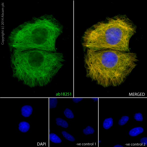 ab18251 staining alpha-Tubulin in Caco-2 cells. The cells were fixed with 100% methanol (5min) and then blocked in 1% BSA/10% normal goat serum/0.3M glycine in 0.1%PBS-Tween for 1h. The cells were then incubated with ab18251 at 5μl/ml and ab7291 at 1µg/ml overnight at +4°C, followed by a further incubation at room temperature for 1h with an anti-rabbit AlexaFluor® 488 (ab150081) at 2 μg/ml (shown in green) and anti-mouse AlexaFluor® 594 (ab150120) at 2 μg/ml (shown in pseudo color red). Nuclear DNA was labelled in blue with DAPI.Negative controls: 1– Rabbit primary antibody and anti-mouse secondary antibody; 2 – Mouse primary antibody and anti-rabbit secondary antibody. Controls 1 and 2 indicate that there is no unspecific reaction between primary and secondary antibodies used.
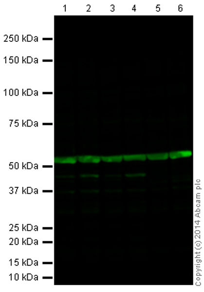 All lanes : Anti-alpha Tubulin antibody (ab18251) at 0.5 µg/mlLane 1 : HeLa (Human epithelial carcinoma cell line) Whole Cell LysateLane 2 : HEK293 (Human embryonic kidney cell line) Whole Cell LysateLane 3 : HepG2 (Human hepatocellular liver carcinoma cell line) Whole Cell LysateLane 4 : Caco 2 (Human colonic carcinoma cell line) Whole Cell LysateLane 5 : NIH 3T3 (Mouse embryonic fibroblast cell line) Whole Cell LysateLane 6 : PC12 (Rat adrenal pheochromocytoma cell line) Whole Cell LysateLysates/proteins at 20 µg per lane.SecondaryGoat Anti-Rabbit IgG H&L (Alexa Fluor® 790) (ab175781) at 1/10000 dilution
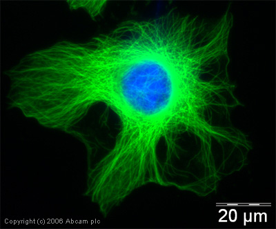
 ab18251 staining alpha Tubulin in the COS7 fibroblast cell line from African Green Monkey by ICC/IF (Immunocytochemistry/immunofluorescence). Cells were fixed with methano and blocked with 5% BSA for 60 minutes at 21°C. Samples were incubated with primary antibody (1/500) for 17 hours at 4°C. An Alexa Fluor®488-conjugated Goat anti-rabbit IgG polyclonal(1/400) was used as the secondary antibody.See Abreview
 ICC/IF image of ab18251 stained human HeLa cells. The cells were methanol fixed (5 min) and incubated with the antibody (ab18251, 1µg/ml) for 1h at room temperature. The secondary antibody (green) was Alexa Fluor® 488 goat anti-rabbit IgG (H+L) used at a 1/1000 dilution for 1h. Image-iTTM FX Signal Enhancer was used as the primary blocking agent, 5% BSA (in TBS-T) was used for all other blocking steps. DAPI was used to stain the cell nuclei (blue).
 All lanes : Anti-alpha Tubulin antibody (ab18251) at 1 µg/mlLane 1 : HeLa (Human epithelial carcinoma cell line) Whole Cell LysateLane 2 : A431 (Human epithelial carcinoma cell line) Whole Cell LysateLysates/proteins at 10 µg per lane.SecondaryGoat Anti-Rabbit IgG H&L (HRP) (ab97051) at 1/50000 dilutiondeveloped using the ECL techniquePerformed under reducing conditions.
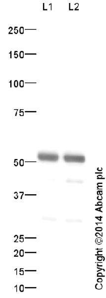
 All lanes : Anti-alpha Tubulin antibody (ab18251) at 0.5 µg/mlLane 1 : HeLa lysateLane 2 : A431 lysateLysates/proteins at 20 µg per lane.SecondaryAlexa Flour Goat polyclonal to Rabbit IgG (700) at 1/10000 dilution
 ab18251 at a 1/8000 dilution staining human HeLa cells by immunocytochemistry. The cells were paraformaldehyde fixed and incubated with the antibody for 30 minutes. The secondary antibody was a Cy3 conjugated Goat Anti-Rabbit IgG (H+L). The image shows staining of an interphase IM cell.This image is courtesy of an Abreview by Kirk McManus submitted on 27 February 2006.See Abreview
 Image courtesy of Human Protein Atlasab18251 staining alpha Tubulin. Paraffin embedded human appendix tissue was incubated with ab18251 (1/2500 dilution) for 30 mins at room temperature. Antigen retrieval was performed by heat induction in citrate buffer pH 6. ab18251 was tested in a tissue microarray (TMA) containing a wide range of normal and cancer tissues as well as a cell microarray consisting of a range of commonly used, well characterised human cell lines. Further results for this antibody can be found at www.proteinatlas.org
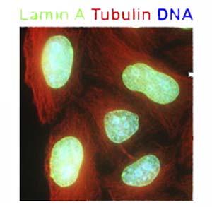 ab18251 staining alpha Tubulin in HeLa cells by Immunocytochemistry/ Immunofluorescence.ab18251 staining alpha Tubulin labeled red.ab20740 staining Lamin A labeled green.Hoechst 33258 staining DNA blue.
Product References
Importance of the CEP215-pericentrin interaction for centrosome maturation during - Importance of the CEP215-pericentrin interaction for centrosome maturation during
Kim S, Rhee K. PLoS One. 2014 Jan 22;9(1):e87016.
ATM specifically mediates repair of double-strand breaks with blocked DNA ends. - ATM specifically mediates repair of double-strand breaks with blocked DNA ends.
Alvarez-Quilon A, Serrano-Benitez A, Lieberman JA, Quintero C, Sanchez-Gutierrez D, Escudero LM, Cortes-Ledesma F. Nat Commun. 2014 Feb 27;5:3347.
Bacillus cereus Certhrax ADP-ribosylates vinculin to disrupt focal adhesion - Bacillus cereus Certhrax ADP-ribosylates vinculin to disrupt focal adhesion
Simon NC, Barbieri JT. J Biol Chem. 2014 Apr 11;289(15):10650-9.
Nuclear motility in glioma cells reveals a cell-line dependent role of various - Nuclear motility in glioma cells reveals a cell-line dependent role of various
Kiss A, Horvath P, Rothballer A, Kutay U, Csucs G. PLoS One. 2014 Apr 1;9(4):e93431.
Cep192 controls the balance of centrosome and non-centrosomal microtubules during - Cep192 controls the balance of centrosome and non-centrosomal microtubules during
O'Rourke BP, Gomez-Ferreria MA, Berk RH, Hackl AM, Nicholas MP, O'Rourke SC, Pelletier L, Sharp DJ. PLoS One. 2014 Jun 27;9(6):e101001.
Beta human papillomavirus E6 expression inhibits stabilization of p53 and - Beta human papillomavirus E6 expression inhibits stabilization of p53 and
Wallace NA, Robinson K, Galloway DA. J Virol. 2014 Jun;88(11):6112-27.
Imaging the alphavirus exit pathway. - Imaging the alphavirus exit pathway.
Martinez MG, Snapp EL, Perumal GS, Macaluso FP, Kielian M. J Virol. 2014 Jun;88(12):6922-33.
Prickle/spiny-legs isoforms control the polarity of the apical microtubule - Prickle/spiny-legs isoforms control the polarity of the apical microtubule
Olofsson J, Sharp KA, Matis M, Cho B, Axelrod JD. Development. 2014 Jul;141(14):2866-74.
Differential sensitivity of Glioma stem cells to Aurora kinase A inhibitors: - Differential sensitivity of Glioma stem cells to Aurora kinase A inhibitors:
Mannino M, Gomez-Roman N, Hochegger H, Chalmers AJ. Stem Cell Res. 2014 Jul;13(1):135-43.
In vitro and in vivo characterization of the actin polymerizing compound - In vitro and in vivo characterization of the actin polymerizing compound
Menhofer MH, Bartel D, Liebl J, Kubisch R, Busse J, Wagner E, Muller R, Vollmar AM, Zahler S. Cardiovasc Res. 2014 Nov 1;104(2):303-14.