![All lanes : Anti-alpha Tubulin (acetyl K40) antibody [EPR16772] (ab179484) at 1/20000 dilutionLane 1 : HeLa (Human epithelial cells from cervix adenocarcinoma) whole cell lysate treated with 500 ng/ml Trichostatin A for 4 hoursLane 2 : Untreated HeLa whole cell lysateLysates/proteins at 10 µg per lane.SecondaryGoat Anti-Rabbit IgG, (H+L), Peroxidase conjugated at 1/1000 dilution](http://www.bioprodhub.com/system/product_images/ab_products/2/sub_1/6029_ab179484-230132-ab179484WB.jpg)
All lanes : Anti-alpha Tubulin (acetyl K40) antibody [EPR16772] (ab179484) at 1/20000 dilutionLane 1 : HeLa (Human epithelial cells from cervix adenocarcinoma) whole cell lysate treated with 500 ng/ml Trichostatin A for 4 hoursLane 2 : Untreated HeLa whole cell lysateLysates/proteins at 10 µg per lane.SecondaryGoat Anti-Rabbit IgG, (H+L), Peroxidase conjugated at 1/1000 dilution
![All lanes : Anti-alpha Tubulin (acetyl K40) antibody [EPR16772] (ab179484) at 1/20000 dilutionLane 1 : C6 (Rat glial tumor cells) whole cell lysate treated with 500 ng/ml Trichostatin A for 4 hoursLane 2 : Untreated C6 whole cell lysateLysates/proteins at 10 µg per lane.SecondaryGoat Anti-Rabbit IgG, (H+L), Peroxidase conjugated at 1/1000 dilution](http://www.bioprodhub.com/system/product_images/ab_products/2/sub_1/6030_ab179484-230133-ab179484WBb.jpg)
All lanes : Anti-alpha Tubulin (acetyl K40) antibody [EPR16772] (ab179484) at 1/20000 dilutionLane 1 : C6 (Rat glial tumor cells) whole cell lysate treated with 500 ng/ml Trichostatin A for 4 hoursLane 2 : Untreated C6 whole cell lysateLysates/proteins at 10 µg per lane.SecondaryGoat Anti-Rabbit IgG, (H+L), Peroxidase conjugated at 1/1000 dilution
![All lanes : Anti-alpha Tubulin (acetyl K40) antibody [EPR16772] (ab179484) at 1/20000 dilutionLane 1 : NIH/3T3 (Mouse embyro fibroblast cells) whole cell lysate treated with 500 ng/ml Trichostatin A for 4 hoursLane 2 : Untreated NIH/3T3 whole cell lysateLysates/proteins at 10 µg per lane.SecondaryLane 1 : Goat Anti-Rabbit IgG, (H+L), Peroxidase conjugated at 1/1000 dilutionLane 2 : Goat Anti-Rabbit IgG, (H+L),Peroxidase conjugated at 1/1000 dilution](http://www.bioprodhub.com/system/product_images/ab_products/2/sub_1/6031_ab179484-230134-ab179484WBc.jpg)
All lanes : Anti-alpha Tubulin (acetyl K40) antibody [EPR16772] (ab179484) at 1/20000 dilutionLane 1 : NIH/3T3 (Mouse embyro fibroblast cells) whole cell lysate treated with 500 ng/ml Trichostatin A for 4 hoursLane 2 : Untreated NIH/3T3 whole cell lysateLysates/proteins at 10 µg per lane.SecondaryLane 1 : Goat Anti-Rabbit IgG, (H+L), Peroxidase conjugated at 1/1000 dilutionLane 2 : Goat Anti-Rabbit IgG, (H+L),Peroxidase conjugated at 1/1000 dilution
![All lanes : Anti-alpha Tubulin (acetyl K40) antibody [EPR16772] (ab179484) at 1/2000 dilutionLane 1 : Mouse brain lysateLane 2 : Mouse kidney lysateLane 3 : Mouse spleen lysateLane 4 : Rat brain lysateLane 5 : Rat heart lysateLane 6 : Human fetal heart lysateLane 7 : Human fetal kidney lysateLysates/proteins at 10 µg per lane.SecondaryGoat Anti-Rabbit IgG, (H+L),Peroxidase conjugated at 1/1000 dilution](http://www.bioprodhub.com/system/product_images/ab_products/2/sub_1/6032_ab179484-230136-ab179484WBd.jpg)
All lanes : Anti-alpha Tubulin (acetyl K40) antibody [EPR16772] (ab179484) at 1/2000 dilutionLane 1 : Mouse brain lysateLane 2 : Mouse kidney lysateLane 3 : Mouse spleen lysateLane 4 : Rat brain lysateLane 5 : Rat heart lysateLane 6 : Human fetal heart lysateLane 7 : Human fetal kidney lysateLysates/proteins at 10 µg per lane.SecondaryGoat Anti-Rabbit IgG, (H+L),Peroxidase conjugated at 1/1000 dilution
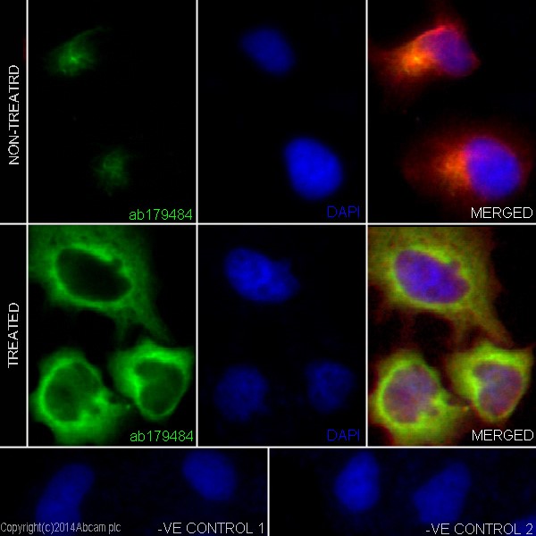
Immunofluorescence analysis of 4% paraformaldehyde fixed, 0.1% tritonX-100 permeabilized HeLa (Human epithelial cells from cervix adenocarcinoma) cells labeling alpha Tubulin (acetyl K40) (green) with ab179484 at 1/500 dilution. Secondary ab: anti-rabbit Alexa Fluor® 488 (ab150077) at 1/200 dilution. Counter stain is labeling tubulin (red) with ab7291 at 1/500 dilution with secondary antibody anti-Mouse AlexaFluor® 594 (ab150120) at 1/500 dilution. Cells are treated with Trichostatin A at 50µg/ml for 4 hours showing cytoplasmic staining. DAPI stains the nucleus in blue. -ve control 1 is ab179484 at 1/500 dilution, ab150120 at 1/500 dilution. -ve control 2 is ab7291 at 1/500 dilution, ab150077 at 1/200 dilution.
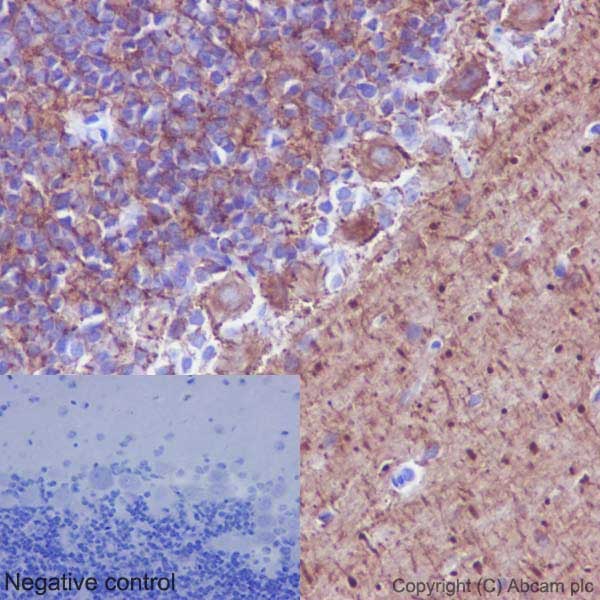
Immunohistochemical analysis of paraffin-embedded Rat cerebellum tissue labeling alpha Tubulin (acetyl K40) with ab179484 at 1/1000 dilution, followed by prediluted HRP Polymer for Rabbit/Mouse IgG. Cytoplasmic staining is observed on Purkinje cells of cerebellum. Counter stained with Hematoxylin.Negative control: Using PBS instead of primary ab, secondary ab is prediluted HRP Polymer for Rabbit/Mouse IgG.
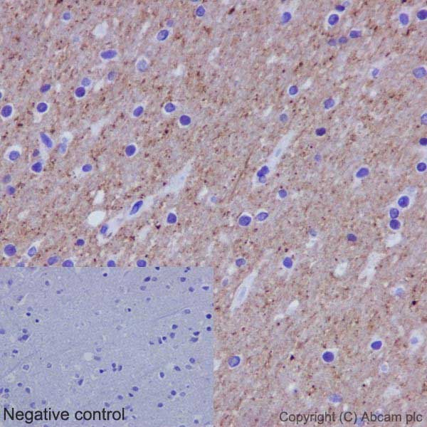
Immunohistochemical analysis of paraffin-embedded Human cerebral cortex tissue labeling alpha Tubulin (acetyl K40) with ab179484 at 1/1000 dilution, followed by prediluted HRP Polymer for Rabbit/Mouse IgG. Cytoplasmic staining is observed on neuron cells of Human brain tissue. Counter stained with Hematoxylin.Negative control: Using PBS instead of primary ab, secondary ab is prediluted HRP Polymer for Rabbit/Mouse IgG.
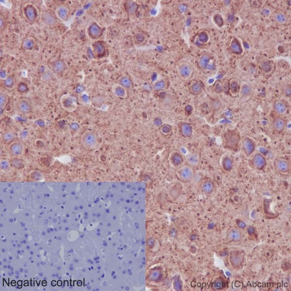
Immunohistochemical analysis of paraffin-embedded Mouse cerebral cortex tissue labeling alpha Tubulin (acetyl K40) with ab179484 at 1/1000 dilution, followed by prediluted HRP Polymer for Rabbit/Mouse IgG. Cytoplasmic staining is observed on neuron cells of Mouse cerebral cortex tissue. Counter stained with Hematoxylin.Negative control: Using PBS instead of primary ab, secondary ab is prediluted HRP Polymer for Rabbit/Mouse IgG.
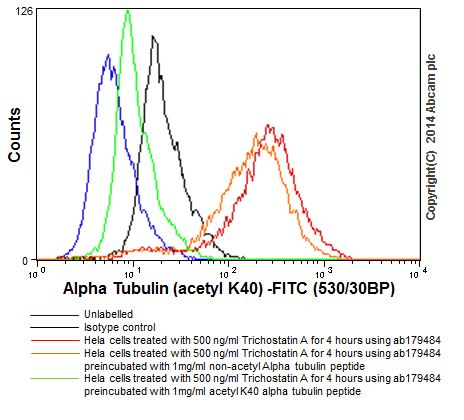
Flow cytometric analysis of 2% paraformaldehyde-fixed HeLa (Human epithelial cells from cervix adenocarcinoma) cells treated with 500 ng/ml Trichostatin A for 4 hours labeling alpha Tubulin (acetyl K40) with ab179484 at 1/240 dilution (red line). Goat anti rabbit IgG (FITC) at 1/150 dilution was used as the secondary antibody. ab179484 preincubated with 1mg/ml acetyl Alpha tubulin (acetyl K40) peptide (green) or non-acetyl Alpha tubulin (acetyl K40) peptide (orange). The isotype control was Rabbit monoclonal IgG (black) and the unlabelled contol was cells without incubation with primary antibody and secondary antibody (blue).
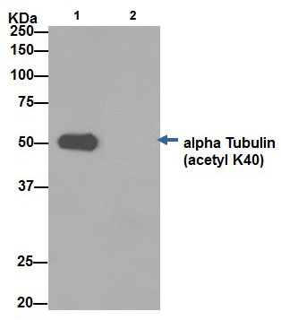
Alpha Tubulin was immunoprecipitated from 1mg of HeLa cells (Human epithelial cells from cervix adenocarcinoma) treated with 500 ng/ml Trichostatin A for 4 hours with ab179484 at 1/70 dilution. Western blot was performed from 10 µg of the immunoprecipitate using ab179484 at 1/1000 dilution. Anti-Rabbit IgG (HRP), specific to the non-reduced form of IgG, was used as secondary antibody at 1/1500 dilution. Left lane: Hela whole cell extract. Right lane: PBS instead of Hela whole cell extract.Blocking and dilution buffer and concentration: 5% NFDM/TBST.
![All lanes : Anti-alpha Tubulin (acetyl K40) antibody [EPR16772] (ab179484) at 1/20000 dilutionLane 1 : HeLa (Human epithelial cells from cervix adenocarcinoma) whole cell lysate treated with 500 ng/ml Trichostatin A for 4 hoursLane 2 : Untreated HeLa whole cell lysateLysates/proteins at 10 µg per lane.SecondaryGoat Anti-Rabbit IgG, (H+L), Peroxidase conjugated at 1/1000 dilution](http://www.bioprodhub.com/system/product_images/ab_products/2/sub_1/6029_ab179484-230132-ab179484WB.jpg)
![All lanes : Anti-alpha Tubulin (acetyl K40) antibody [EPR16772] (ab179484) at 1/20000 dilutionLane 1 : C6 (Rat glial tumor cells) whole cell lysate treated with 500 ng/ml Trichostatin A for 4 hoursLane 2 : Untreated C6 whole cell lysateLysates/proteins at 10 µg per lane.SecondaryGoat Anti-Rabbit IgG, (H+L), Peroxidase conjugated at 1/1000 dilution](http://www.bioprodhub.com/system/product_images/ab_products/2/sub_1/6030_ab179484-230133-ab179484WBb.jpg)
![All lanes : Anti-alpha Tubulin (acetyl K40) antibody [EPR16772] (ab179484) at 1/20000 dilutionLane 1 : NIH/3T3 (Mouse embyro fibroblast cells) whole cell lysate treated with 500 ng/ml Trichostatin A for 4 hoursLane 2 : Untreated NIH/3T3 whole cell lysateLysates/proteins at 10 µg per lane.SecondaryLane 1 : Goat Anti-Rabbit IgG, (H+L), Peroxidase conjugated at 1/1000 dilutionLane 2 : Goat Anti-Rabbit IgG, (H+L),Peroxidase conjugated at 1/1000 dilution](http://www.bioprodhub.com/system/product_images/ab_products/2/sub_1/6031_ab179484-230134-ab179484WBc.jpg)
![All lanes : Anti-alpha Tubulin (acetyl K40) antibody [EPR16772] (ab179484) at 1/2000 dilutionLane 1 : Mouse brain lysateLane 2 : Mouse kidney lysateLane 3 : Mouse spleen lysateLane 4 : Rat brain lysateLane 5 : Rat heart lysateLane 6 : Human fetal heart lysateLane 7 : Human fetal kidney lysateLysates/proteins at 10 µg per lane.SecondaryGoat Anti-Rabbit IgG, (H+L),Peroxidase conjugated at 1/1000 dilution](http://www.bioprodhub.com/system/product_images/ab_products/2/sub_1/6032_ab179484-230136-ab179484WBd.jpg)


