Anti-alpha Adaptin antibody [AP6]
| Name | Anti-alpha Adaptin antibody [AP6] |
|---|---|
| Supplier | Abcam |
| Catalog | ab2730 |
| Prices | $403.00 |
| Sizes | 100 µl |
| Host | Mouse |
| Clonality | Monoclonal |
| Isotype | IgG1 |
| Clone | AP6 |
| Applications | ICC/IF ICC/IF ICC/IF B/N Electron microscopy ELISA B/N IP WB IHC-F FC |
| Species Reactivities | Mouse, Rat, Hamster, Bovine, Human, Drosophila, Primate |
| Antigen | Other Immunogen Type corresponding to alpha Adaptin |
| Description | Mouse Monoclonal |
| Gene | AP2A2 |
| Conjugate | Unconjugated |
| Supplier Page | Shop |
Product images
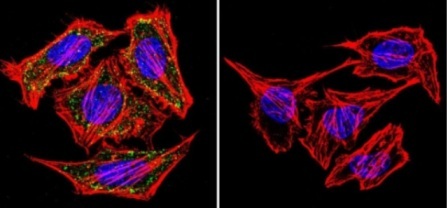
 Immunofluorescent analysis of alpha Adaptin using alpha Adaptin Monoclonal Antibody (AP6) (ab2730) shows staining in U251 Cells. alpha Adaptin (green) F-Actin staining with Phalloidin (red) and nuclei with DAPI (blue) is shown. Cells were grown on chamber slides and fixed with formaldehyde prior to staining. Cells were probed without (control) or with an antibody recognizing alpha Adaptin (ab2730) at a dilution of 1:20 over night at 4C and incubated with a DyLight-488 conjugated secondary antibody. Images were taken at 60X magnification.
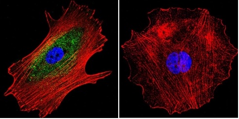 Immunofluorescent analysis of alpha Adaptin using alpha Adaptin Monoclonal Antibody (AP6) (ab2720) shows staining in MCF-7 Cells. alpha Adaptin (green) F-Actin staining with Phalloidin (red) and nuclei with DAPI (blue) is shown. Cells were grown on chamber slides and fixed with formaldehyde prior to staining. Cells were probed without (control) or with an antibody recognizing alpha Adaptin (ab2730) at a dilution of 1:20 over night at 4 C and incubated with a DyLight-488 conjugated secondary antibody. Images were taken at 60X magnification.
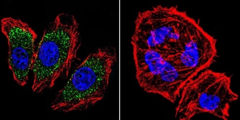
 Immunofluorescent analysis of alpha Adaptin using alpha Adaptin Monoclonal Antibody (AP6) (ab2730) shows staining in Hela Cells. alpha Adaptin (green) F-Actin staining with Phalloidin (red) and nuclei with DAPI (blue) is shown. Cells were grown on chamber slides and fixed with formaldehyde prior to staining. Cells were probed without (control) or with an antibody recognizing alpha Adaptin (ab2720) at a dilution of 1:20 over night at 4 C and incubated with a DyLight-488 conjugated secondary antibody. Images were taken at 60X magnification.
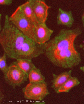 ICC/IF image of ab2730 stained MCF7 cells. The cells were 4% formaldehyde fixed (10 min) and then incubated in 1%BSA / 10% normal goat serum / 0.3M glycine in 0.1% PBS-Tween for 1h to permeabilise the cells and block non-specific protein-protein interactions. The cells were then incubated with the antibody (ab2730, 5µg/ml) overnight at +4°C. The secondary antibody (green) was Alexa Fluor® 488 goat anti-mouse IgG (H+L) used at a 1/1000 dilution for 1h. Alexa Fluor® 594 WGA was used to label plasma membranes (red) at a 1/200 dilution for 1h. DAPI was used to stain the cell nuclei (blue) at a concentration of 1.43µM.
![Overlay histogram showing MCF7 cells stained with ab2730 (red line). The cells were fixed with 4% paraformaldehyde (10 min) and then permeabilized with 0.1% PBS-Tween for 20 min. The cells were then incubated in 1x PBS / 10% normal goat serum / 0.3M glycine to block non-specific protein-protein interactions followed by the antibody (ab2730, 1µg/1x106 cells) for 30 min at 22ºC. The secondary antibody used was a goat anti-mouse DyLight® 488 (IgG; H+L) (ab96879) at 1/500 dilution for 30 min at 22ºC. Isotype control antibody (black line) was mouse IgG1 [ICIGG1] (ab91353, 2µg/1x106 cells) used under the same conditions. Acquisition of >5,000 events was performed.](http://www.bioprodhub.com/system/product_images/ab_products/2/sub_1/5626_alpha-Adaptin-Primary-antibodies-ab2730-3.jpg) Overlay histogram showing MCF7 cells stained with ab2730 (red line). The cells were fixed with 4% paraformaldehyde (10 min) and then permeabilized with 0.1% PBS-Tween for 20 min. The cells were then incubated in 1x PBS / 10% normal goat serum / 0.3M glycine to block non-specific protein-protein interactions followed by the antibody (ab2730, 1µg/1x106 cells) for 30 min at 22ºC. The secondary antibody used was a goat anti-mouse DyLight® 488 (IgG; H+L) (ab96879) at 1/500 dilution for 30 min at 22ºC. Isotype control antibody (black line) was mouse IgG1 [ICIGG1] (ab91353, 2µg/1x106 cells) used under the same conditions. Acquisition of >5,000 events was performed.
Overlay histogram showing MCF7 cells stained with ab2730 (red line). The cells were fixed with 4% paraformaldehyde (10 min) and then permeabilized with 0.1% PBS-Tween for 20 min. The cells were then incubated in 1x PBS / 10% normal goat serum / 0.3M glycine to block non-specific protein-protein interactions followed by the antibody (ab2730, 1µg/1x106 cells) for 30 min at 22ºC. The secondary antibody used was a goat anti-mouse DyLight® 488 (IgG; H+L) (ab96879) at 1/500 dilution for 30 min at 22ºC. Isotype control antibody (black line) was mouse IgG1 [ICIGG1] (ab91353, 2µg/1x106 cells) used under the same conditions. Acquisition of >5,000 events was performed.
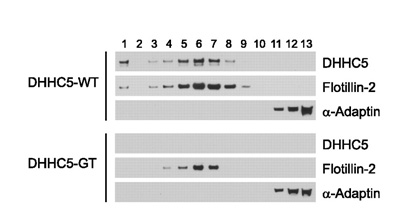 Image from Li Y et al, J Biol Chem. 2012 Jan 2;287(1):523-30. Epub 2011 Nov 11, Fig 2. DOI 10.1074/jbc.M111.306183 January 2, 2012 The Journal of Biological Chemistry, 287, 523-530.ab2730 used in Western Blot at a 1/1000 dilution.Sucrose density gradient fractionation of detergent resistant cell lysates from WT and DHHC5-GT murine neuronal stem cells. The isolation of detergent-resistant membranes was performed according to a previously published protocol. NSCs growing in growth medium (~300 µl of packed cells) were harvested and washed three times with ice-cold PBS. The cells were gently resuspended by pipetting up and down in 700 µl of 1% Triton X-100 in HEPES buffer (25 mm HEPES-HCl, pH 6.5, 150 mm NaCl, 1 mm EDTA, and protease inhibitor mixture), homogenized using a Teflon-coated Dounce homogenizer (30 strokes), then incubated on ice for 30 min, and fractionated on a sucrose density gradient. The fractions were collected from top (fraction 1) to bottom (fraction 13) and analyzed by
Product References
Cdc42 and the Rho GEF intersectin-1 collaborate with Nck to promote - Cdc42 and the Rho GEF intersectin-1 collaborate with Nck to promote
Humphries AC, Donnelly SK, Way M. J Cell Sci. 2014 Feb 1;127(Pt 3):673-85.
Actin and dynamin2 dynamics and interplay during clathrin-mediated endocytosis. - Actin and dynamin2 dynamics and interplay during clathrin-mediated endocytosis.
Grassart A, Cheng AT, Hong SH, Zhang F, Zenzer N, Feng Y, Briner DM, Davis GD, Malkov D, Drubin DG. J Cell Biol. 2014 Jun 9;205(5):721-35.
Cis and trans regulatory mechanisms control AP2-mediated B cell receptor - Cis and trans regulatory mechanisms control AP2-mediated B cell receptor
Busman-Sahay K, Drake L, Sitaram A, Marks M, Drake JR. PLoS One. 2013;8(1):e54938.
DHHC5 protein palmitoylates flotillin-2 and is rapidly degraded on induction of - DHHC5 protein palmitoylates flotillin-2 and is rapidly degraded on induction of
Li Y, Martin BR, Cravatt BF, Hofmann SL. J Biol Chem. 2012 Jan 2;287(1):523-30.
Endocytosis regulates cell soma translocation and the distribution of adhesion - Endocytosis regulates cell soma translocation and the distribution of adhesion
Shieh JC, Schaar BT, Srinivasan K, Brodsky FM, McConnell SK. PLoS One. 2011 Mar 22;6(3):e17802.
Crystal structure of nucleotide-free dynamin. - Crystal structure of nucleotide-free dynamin.
Faelber K, Posor Y, Gao S, Held M, Roske Y, Schulze D, Haucke V, Noe F, Daumke O. Nature. 2011 Sep 18;477(7366):556-60.
Clathrin mediates integrin endocytosis for focal adhesion disassembly in - Clathrin mediates integrin endocytosis for focal adhesion disassembly in
Ezratty EJ, Bertaux C, Marcantonio EE, Gundersen GG. J Cell Biol. 2009 Nov 30;187(5):733-47.
Molecular basis for the sorting of the SNARE VAMP7 into endocytic clathrin-coated - Molecular basis for the sorting of the SNARE VAMP7 into endocytic clathrin-coated
Pryor PR, Jackson L, Gray SR, Edeling MA, Thompson A, Sanderson CM, Evans PR, Owen DJ, Luzio JP. Cell. 2008 Sep 5;134(5):817-27.
Rho/ROCK and myosin II control the polarized distribution of endocytic clathrin - Rho/ROCK and myosin II control the polarized distribution of endocytic clathrin
Samaniego R, Sanchez-Martin L, Estecha A, Sanchez-Mateos P. J Cell Sci. 2007 Oct 15;120(Pt 20):3534-43. Epub 2007 Sep 25.
Transmitter release face Ca2+ channel clusters persist at isolated presynaptic - Transmitter release face Ca2+ channel clusters persist at isolated presynaptic
Sun L, Li Q, Khanna R, Chan AW, Wong F, Stanley EF. Eur J Neurosci. 2006 Mar;23(5):1391-6.