Anti-alpha Adaptin antibody [AC1-M11]
| Name | Anti-alpha Adaptin antibody [AC1-M11] |
|---|---|
| Supplier | Abcam |
| Catalog | ab2807 |
| Prices | $388.00 |
| Sizes | 500 µl |
| Host | Mouse |
| Clonality | Monoclonal |
| Isotype | IgG2a |
| Clone | AC1-M11 |
| Applications | IP WB ICC/IF ICC/IF ELISA IHC-P FC |
| Species Reactivities | Mouse, Rat, Chicken, Bovine, Human, Pig, Primate, Amphibians |
| Antigen | Full length native protein (purified) corresponding to Cow alpha Adaptin |
| Description | Mouse Monoclonal |
| Gene | AP2A2 |
| Conjugate | Unconjugated |
| Supplier Page | Shop |
Product images
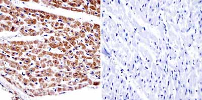 Immunohistochemistry was performed on normal biopsies of deparaffinized Human heart tissue. To expose target proteins heat induced antigen retrieval was performed using 10mM sodium citrate (pH6.0) buffer and microwaved for 8-15 minutes. Following antigen retrieval tissues were blocked in 3% BSA-PBS for 30 minutes at room temperature. Tissues were then probed at a dilution of 1:20 with a Mouse monoclonal antibody recognizing alpha Adaptin (ab2807) or without primary antibody (negative control) overnight at 4�C in a humidified chamber. Tissues were washed extensively with PBST and endogenous peroxidase activity was quenched with a peroxidase suppressor. Detection was performed using a biotin-conjugated secondary antibody and SA-HRP followed by colorimetric detection using DAB. Tissues were counterstained with hematoxylin and prepped for mounting.
 Immunolocalization of alpha-adaptin in NRK cells using ab2807.
 Immunolocalization of alpha-adaptin in NRK cells using ab2807 (low power image of image1).
![Anti-alpha Adaptin antibody [AC1-M11] (ab2807) at 1/500 dilution + Lysate was prepared from mouse neuroblastoma cells at 10 µgSecondaryHRP-conjugated donkey monoclonal to mouse IgG at 1/1000 dilutiondeveloped using the ECL techniquePerformed under reducing conditions.Observed band size : 100 kDa (why is the actual band size different from the predicted?)Exposure time : 10 secondsThis image is a courtesy of Anonymous AbreviewSee Abreview](http://www.bioprodhub.com/system/product_images/ab_products/2/sub_1/5620_alpha-Adaptin-Primary-antibodies-ab2807-2.jpg) Anti-alpha Adaptin antibody [AC1-M11] (ab2807) at 1/500 dilution + Lysate was prepared from mouse neuroblastoma cells at 10 µgSecondaryHRP-conjugated donkey monoclonal to mouse IgG at 1/1000 dilutiondeveloped using the ECL techniquePerformed under reducing conditions.Observed band size : 100 kDa (why is the actual band size different from the predicted?)Exposure time : 10 secondsThis image is a courtesy of Anonymous AbreviewSee Abreview
Anti-alpha Adaptin antibody [AC1-M11] (ab2807) at 1/500 dilution + Lysate was prepared from mouse neuroblastoma cells at 10 µgSecondaryHRP-conjugated donkey monoclonal to mouse IgG at 1/1000 dilutiondeveloped using the ECL techniquePerformed under reducing conditions.Observed band size : 100 kDa (why is the actual band size different from the predicted?)Exposure time : 10 secondsThis image is a courtesy of Anonymous AbreviewSee Abreview
![Overlay histogram showing HepG2 cells stained with ab2807 (red line). The cells were fixed with 80% methanol (5 min) and then permeabilized with 0.1% PBS-Tween for 20 min. The cells were then incubated in 1x PBS / 10% normal goat serum / 0.3M glycine to block non-specific protein-protein interactions followed by the antibody (ab2807, 1/100 dilution) for 30 min at 22ºC. The secondary antibody used was DyLight® 488 goat anti-mouse IgG (H+L) (ab96879) at 1/500 dilution for 30 min at 22ºC. Isotype control antibody (black line) was mouse IgG2a [ICIGG2A] (ab91361, 2µg/1x106 cells) used under the same conditions. Acquisition of >5,000 events was performed. This antibody gave a positive signal in HepG2 cells fixed with 4% paraformaldehyde (10 min)/permeabilized with 0.1% PBS-Tween for 20 min used under the same conditions.](http://www.bioprodhub.com/system/product_images/ab_products/2/sub_1/5621_alpha-Adaptin-Primary-antibodies-ab2807-3.jpg) Overlay histogram showing HepG2 cells stained with ab2807 (red line). The cells were fixed with 80% methanol (5 min) and then permeabilized with 0.1% PBS-Tween for 20 min. The cells were then incubated in 1x PBS / 10% normal goat serum / 0.3M glycine to block non-specific protein-protein interactions followed by the antibody (ab2807, 1/100 dilution) for 30 min at 22ºC. The secondary antibody used was DyLight® 488 goat anti-mouse IgG (H+L) (ab96879) at 1/500 dilution for 30 min at 22ºC. Isotype control antibody (black line) was mouse IgG2a [ICIGG2A] (ab91361, 2µg/1x106 cells) used under the same conditions. Acquisition of >5,000 events was performed. This antibody gave a positive signal in HepG2 cells fixed with 4% paraformaldehyde (10 min)/permeabilized with 0.1% PBS-Tween for 20 min used under the same conditions.
Overlay histogram showing HepG2 cells stained with ab2807 (red line). The cells were fixed with 80% methanol (5 min) and then permeabilized with 0.1% PBS-Tween for 20 min. The cells were then incubated in 1x PBS / 10% normal goat serum / 0.3M glycine to block non-specific protein-protein interactions followed by the antibody (ab2807, 1/100 dilution) for 30 min at 22ºC. The secondary antibody used was DyLight® 488 goat anti-mouse IgG (H+L) (ab96879) at 1/500 dilution for 30 min at 22ºC. Isotype control antibody (black line) was mouse IgG2a [ICIGG2A] (ab91361, 2µg/1x106 cells) used under the same conditions. Acquisition of >5,000 events was performed. This antibody gave a positive signal in HepG2 cells fixed with 4% paraformaldehyde (10 min)/permeabilized with 0.1% PBS-Tween for 20 min used under the same conditions.
Product References
Clathrin is required for Scar/Wave-mediated lamellipodium formation. - Clathrin is required for Scar/Wave-mediated lamellipodium formation.
Gautier JJ, Lomakina ME, Bouslama-Oueghlani L, Derivery E, Beilinson H, Faigle W, Loew D, Louvard D, Echard A, Alexandrova AY, Baum B, Gautreau A. J Cell Sci. 2011 Oct 15;124(Pt 20):3414-27.
100-kD coated vesicle proteins: molecular heterogeneity and intracellular - 100-kD coated vesicle proteins: molecular heterogeneity and intracellular
Robinson MS. J Cell Biol. 1987 Apr;104(4):887-95.