Anti-alpha 1 Antitrypsin antibody
| Name | Anti-alpha 1 Antitrypsin antibody |
|---|---|
| Supplier | Abcam |
| Catalog | ab17438 |
| Prices | $378.00 |
| Sizes | 250 µg |
| Host | Chicken |
| Clonality | Polyclonal |
| Isotype | IgY |
| Applications | WB ELISA ICC/IF ICC/IF |
| Species Reactivities | Human |
| Antigen | Synthetic peptide: DAAQKTDTSHHD , corresponding to amino acids 30-41 of Human alpha 1 Antitrypsin Synthetic peptide: ITKFLNEDRRS, corresponding to amino acids 296-307 of Human alpha 1 Antitrypsin Run BLAST with Run BLAST with Run BLAST with Run BLAST with |
| Description | Chicken Polyclonal |
| Gene | SERPINA1 |
| Conjugate | Unconjugated |
| Supplier Page | Shop |
Product images
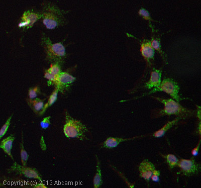 ICC/IF image of ab17438 stained HepG2 cells. The cells were 4% paraformaldehyde fixed (10 min) and then incubated in 1%BSA / 10% normal goat serum / 0.3M glycine in 0.1% PBS-Tween for 1h to permeabilise the cells and block non-specific protein-protein interactions. The cells were then incubated with the antibody (ab17438, 5µg/ml) overnight at +4°C. The secondary antibody (green) was ab96947, DyLight® 488 goat anti-chicken IgG (H+L) used at a 1/250 dilution for 1h. Alexa Fluor® 594 WGA was used to label plasma membranes (red) at a 1/200 dilution for 1h. DAPI was used to stain the cell nuclei (blue) at a concentration of 1.43µM.