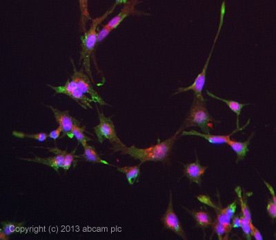
ICC/IF image of ab126820 stained HepG2 cells. The cells were 4% formaldehyde fixed (10 min) and then incubated in 1%BSA / 10% normal goat serum / 0.3M glycine in 0.1% PBS-Tween for 1h to permeabilise the cells and block non-specific protein-protein interactions. The cells were then incubated with the antibody ab126820 at 1/100 dilution overnight at +4°C. The secondary antibody (green) was DyLight® 488 goat anti- mouse (ab96879) IgG (H+L) used at a 1/250 dilution for 1h. Alexa Fluor® 594 WGA was used to label plasma membranes (red) at a 1/200 dilution for 1h. DAPI was used to stain the cell nuclei (blue) at a concentration of 1.43µM. This antibody also gave a positive result in methanol fixed (100%, 5min) hepG2 cells used at 1/100 dilution.
![Anti-Alkaline Phosphatase, Tissue Non-Specific antibody [2F4] (ab126820) at 1/500 dilution + Recombinant Human protein ALPL (AA: 18-502)](http://www.bioprodhub.com/system/product_images/ab_products/2/sub_1/5096_Alkaline-Phosphatase-Tissue-Non-Specific-Primary-antibodies-ab126820-1.jpg)
Anti-Alkaline Phosphatase, Tissue Non-Specific antibody [2F4] (ab126820) at 1/500 dilution + Recombinant Human protein ALPL (AA: 18-502)
![All lanes : Anti-Alkaline Phosphatase, Tissue Non-Specific antibody [2F4] (ab126820) at 1/500 dilutionLane 1 : HeLa cell lysateLane 2 : NTERA-2 cell lysate](http://www.bioprodhub.com/system/product_images/ab_products/2/sub_1/5097_Alkaline-Phosphatase-Tissue-Non-Specific-Primary-antibodies-ab126820-2.jpg)
All lanes : Anti-Alkaline Phosphatase, Tissue Non-Specific antibody [2F4] (ab126820) at 1/500 dilutionLane 1 : HeLa cell lysateLane 2 : NTERA-2 cell lysate
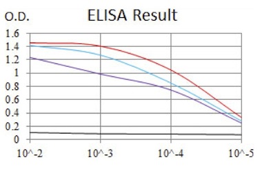
Example of standard curves obtained using ab126820. Control antigen 100ng (black), antigen at 10ng (purple), antigen at 50ng (blue), antigen at 100ng (red)
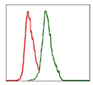
Flow cytometric analysis of MCF-7 cells using ab126820 at 1/200 (green) and negative control (red).
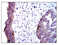
ab126820, at 1/200, staining Alkaline Phosphatase, Tissue Non-Specific in paraffin embedded Human ovarian cancer tissues by Immunohistochemistry with DAB staining.