![Anti-AKR1B10 antibody [EPR14421] (ab192865) at 1/10000 dilution + A549 cell lysate at 20 µgSecondaryGoat Anti-Rabbit IgG, (H+L), Peroxidase conjugated at 1/1000 dilution](http://www.bioprodhub.com/system/product_images/ab_products/2/sub_1/4189_ab192865-230667-ab1928651.jpg)
Anti-AKR1B10 antibody [EPR14421] (ab192865) at 1/10000 dilution + A549 cell lysate at 20 µgSecondaryGoat Anti-Rabbit IgG, (H+L), Peroxidase conjugated at 1/1000 dilution
![Anti-AKR1B10 antibody [EPR14421] (ab192865) at 1/1000 dilution + SW480 cell lysate at 20 µgSecondaryGoat Anti-Rabbit IgG, (H+L), Peroxidase conjugated at 1/1000 dilution](http://www.bioprodhub.com/system/product_images/ab_products/2/sub_1/4190_ab192865-230666-ab1928652.jpg)
Anti-AKR1B10 antibody [EPR14421] (ab192865) at 1/1000 dilution + SW480 cell lysate at 20 µgSecondaryGoat Anti-Rabbit IgG, (H+L), Peroxidase conjugated at 1/1000 dilution
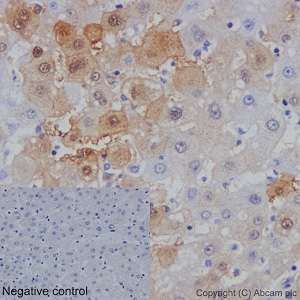
Immunohistochemical analysis of paraffin embedded Human liver tissue labeling AKR1B10 with ab192865 at a 1/1000 dilution. A prediluted HRP Polymer for Rabbit IgG was used as the secondary antibody. Hematoxylin counterstain.
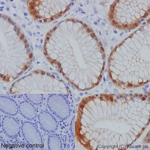
Immunohistochemical analysis of paraffin embedded human stomach tissue sections labeling AKR1B10 using ab192865 at a 1/1000 dilution. A prediluted HRP Polymer for Rabbit IgG was used as the secondary antibody. Hematoxylin counterstain.

Immunofluorescent analysis of 4% paraformaldehyde fixed SW480 cells labeling AKR1B10 with ab192865 at a 1/50 dilution. A Goat anti rabbit IgG (Alexa Fluor®488) (ab150077) was used as the secondary antibody at a 1/400 dilution. Counterstain DAPI. Cells were permeabilized using 0.1% Triton X-100.
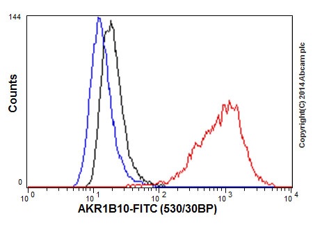
Flow cytometric analysis of A549 cells labeling AKR1B10 using ab192865 at a 1/50 dilution (red). Goat anti rabbit IgG (FITC) was used as the secondary antibody at a 1/150 dilution. Isotype control Rabbit monoclonal IgG (black). Unlabeled control cells without incubation with primary antibody and secondary antibody (blue). Cells were fixed in 2% paraformaldehyde.